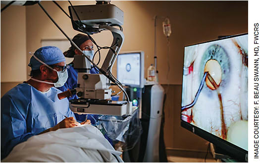
Upon embarking on my surgical fellowship 6 years ago, I, like many of my peers, was entrenched in traditional surgical practices. Operating with shoes off, peering through oculars, employing a peristaltic vacuum phacoemulsification platform, and utilizing large, what some would call “normal” incisions characterized my approach. I had never encountered femtosecond laser-assisted cataract surgery (FLACS), venturi-powered phacoemulsification or 3D heads-up visualization.
However, within a year of joining a forward-thinking, progressive practice, everything changed. The era of conventionality ended, replaced by a new frontier of surgical innovation. I found myself regularly performing FLACS through 3D heads-up visualization, all while wearing my shoes and employing a venturi vacuum phacoemulsification system. Needless to say, the learning curve for mastering these innovations — particularly in the realm of 3D heads-up surgery — was steep.
Initially foreign and uncomfortable, 3D heads-up surgery soon became second nature through deliberate practice and quickly proved worthwhile. The transition was instrumental in enhancing my surgical skills. This innovative approach has delivered unparalleled visualization capabilities, allowing me to identify and navigate previously challenging aspects of surgery. The presence of my mentors, who could see exactly what I saw directly off of my back shoulder, provided invaluable guidance, steering me towards success while averting potential complications.
Moreover, the freedom to adopt any posture during surgery added comfort to my practice. What truly stands out is how this technology involves every member of the operating room (OR) team. This seemingly minor benefit not only sparked enthusiasm for ocular surgery among staff but also enhanced their efficiency and knowledge.
ENHANCED VISUALIZATION
Superior visualization is a cornerstone of heads-up surgery. Not only does it provide a larger image, but also delivers higher-definition visuals through the computer’s processor. Ophthalmic surgery involves subtle nuances that demand constant mastery. With heads-up surgery, identifying subtle changes in lens behavior or recognizing missing zonules becomes a reality. Also, losing the capsulorhexis, once a frustrating occurrence, became a matter of poor visualization rather than surgical skill. With 3D heads-up surgery, I could zoom in on problematic areas, focus my attention and regain control over the procedure.
Recent software iterations facilitate the display of patient data, which includes IOL calculations and keratometry readings in real-time during surgery, along with intraoperative aberrometry in a picture-in-picture format, eliminating the need to divert attention from the screen. Filters are also available to visualize blood or, for retina surgeons, identify the internal limiting membrane. The various companies producing heads-up systems allow for customization of hue, brightness and saturation, ensuring pristine visualization upon installation in your practice.
PRACTICAL IMPLEMENTATION
Another concern associated with the adoption of 3D heads-up technology pertains to its implementation and the potential impact on OR spatial dynamics, efficiency and maintenance requirements. I have encountered two distinct models of implementation.
The first model involves the use of a stationary screen within the OR, where the patient is positioned either feet first or head first, depending on the operative eye. This approach is highly efficient and necessitates the relocation of the phaco machine as needed. In my practice, I prioritize the sequencing of left-eye surgeries followed by right-eye surgeries to streamline this process.
The second model entails a complete system repositioning to the opposite side of the operating room. While eye laterality remains a consideration in this model, it demands the transfer of all associated equipment. The feasibility of either system largely hinges on the capabilities of the anesthesia machines in use. It is worth noting that larger anesthesia machines may require the use of the second system, which can be cumbersome. The incorporation of small bed monitors plays a pivotal role in successfully implementing the first system, avoiding dependence on the anesthesia machine’s location.

Maintenance costs for these 3D heads-up systems are in line with typical ophthalmic equipment maintenance expenses. In my practice, we allocate approximately $2,200 annually to cover maintenance costs associated with our 3D heads-up system.
ADVANCED RECORDING FEATURES
One of the remarkable features inherent in most heads-up visualization systems is their integrated recording capabilities, which contribute significantly to the enhancement of surgical practices. These systems have the capacity to capture surgeries in stunning 3D quality, preserving the precise visual context experienced during the actual procedure. Furthermore, these recorded surgeries can be seamlessly converted into 2D video formats, thereby accommodating the current presentation landscape where the integration of 3D video remains somewhat limited within professional settings.
The utility of these recording capabilities extends beyond mere documentation; they serve as an invaluable tool for continuous skill refinement, akin to the process of reviewing game film in sports. By revisiting these recorded surgeries, surgeons have the opportunity to engage in critical self-assessment, scrutinizing their techniques, decision-making and overall performance. This not only allows for the identification of areas requiring improvement but also fosters a culture of continuous learning and refinement. The playback quality of these recordings is exceptional.
ENHANCING EFFICIENCY
The impact of 3D heads-up surgery extends to the efficiency of my staff. Previously, most of them had only observed cataract surgeries on low-resolution two-dimensional monitors, leading to boredom and poor understanding. Now, they are actively engaged, closely monitoring the progress of the procedure in real-time. This newfound awareness allows them to prepare for upcoming tasks, optimize patient flow and anticipate potential challenges. When complications arise, my staff can identify them before I even utter a word, sharpening their focus and readiness. The vitrectomy set, backup IOLs and other necessary equipment are already being opened and primed, reducing stress in an already stressful situation.
I am fortunate to have an excellent staff that is very engaged in what is happening and how they can help. I remember a recent case where everything seemed to be working against me. My staff was able to see that I was struggling creating an urgency to help. Every eye and mind in that room was focused on the patient. I called for materials and instruments that I do not commonly use, and they were made available quickly. I credit 3D heads-up visualization for creating a big share of the staff’s new interest and for facilitating the anticipation of how they can be of better assistance.
A TEACHING TOOL FOR THE FUTURE ... OR NOW!
In addition to facilitating greater staff efficiency, 3D heads-up visualization serves as an invaluable tool for teaching staff as well as medical students, residents, fellows and optometrists. I remember my time as a resident, observing cataract surgery on a two-dimensional screen, unable to appreciate the depth and intricacies of the procedure. Even when granted a seat at the operating microscope, I was limited to 2D visualization as the observer oculars only pull from one lens.
I believe this limitation is a major contributor to complications early in a surgeon’s career. Residents and fellows may be training with the most brilliant of surgeons and miss some of the most important surgical wisdom they have to impart. With heads-up surgery, the paradigm shifts, fostering greater understanding from an earlier stage, which increases patient safety.
IMPROVED SURGEON HEALTH
The pursuit of a long and successful career in ophthalmology is a paramount concern for every surgeon. Beyond the mastery of surgical skills and the delivery of exceptional patient care, sustaining peak physical and mental condition is essential. Heads-up surgery emerges as a transformative asset in achieving these goals — that’s a benefit that cannot be overstated.

Traditionally, surgeons using conventional microscopes must adapt their posture to align with the microscope’s eyepiece or oculars. This requirement places considerable strain on the neck and back, as surgeons are compelled to maintain a specific posture throughout the duration of the procedure. Taller surgeons often find themselves hunched over, peering down at the oculars, while their shorter counterparts must stretch and extend their necks to gaze upwards. This consistent physical strain, endured over years of practice, can take a significant toll on surgeons’ musculoskeletal health and even curtail their careers, as confirmed by a 2020 survey conducted by Schechel et al.
Herein lies one of the transformative advantages of heads-up visualization: Heads-up systems empower surgeons to operate comfortably in virtually any posture they choose, irrespective of their height or the position of the patient. The reduction in neck and back strain translates into not only improved surgeon well-being but also optimized performance. The ability to sustain peak physical condition over the course of a demanding career is essential for maintaining the highest standards of care for patients.
VALUE BEYOND COST
So, with these benefits, why would surgeons not adopt 3D heads-up visualization? One common objection is the perceived high cost. Some surgeons argue that they operate effectively with oculars and question the justification for investing in this technology. Currently priced between $45,000 and $450,000, 3D systems encompass a 3D microscope camera, a large display (55 to 65 inches), and the necessary computer, with the added challenge that it is not billable.
While these concerns are valid, I have experienced substantial value-added benefits to my practice that extend beyond immediate monetary returns. The intangible benefits quickly manifest as referrals, triggered by word-of-mouth recommendations from OR staff who can now witness the precision of my surgeries. In my multi-specialty ASC, surgeons from various disciplines have started referring patients due to their newfound understanding of my procedures. Additionally, I have attracted more referrals from optometrists by inviting them to observe live surgeries in 3D, setting myself apart and piquing their interest in the process.
CONCLUSION
3D heads-up surgery has transcended its initial perception as a mere novelty and has emerged as an indispensable tool in my practice. The benefits it offers in terms of improved surgical skills, staff efficiency, surgeon well-being, recording capabilities and enhanced teaching capabilities far outweigh the initial cost concerns.
As technology continues to advance, the field of ophthalmology stands to gain even greater insights and improvements from this innovative approach. It is imperative that we embrace these advances to provide the best possible care to our patients and prepare the next generation of surgeons for success. OM


