Peanut butter and chocolate. Chicken and waffles. Watermelon and salt.
Each ingredient tastes good enough on its own. But put them together and you get a flavor combo that delights the taste buds.
So it is with corneal tomography and topography: Each modality stands well on its own. But put them together and they form a veritable power couple when it comes to evaluating patients for refractive surgery and spotting dry eye disease, keratoconus and other ophthalmic villains.
“Corneal tomography and topography should not be exclusive. Any patient who is getting corneal tomography should almost certainly have topography, as well,” says Arjan Hura, MD, a cataract, refractive and anterior segment surgeon at the Maloney-Shamie Vision Institute in Los Angeles. “I get topography and tomography on every single cataract and refractive surgery patient.”
Dr. Hura is not alone. Many ophthalmologists recognize the unique strengths of each modality that give both a place at the table of corneal imaging.
TOPOGRAPHY SYSTEMS
Below is a listing of devices that offer topography, listed by manufacturer.
Alcon
Wavelight Topolyzer Vario
myalcon.com/professional/
Cassini Technologies
Cassini
https://cassini-technologies.com
Marco/Nidek
OPD-Scan III Wavefront Aberrometer
OPD-Scan III Visual System
https://marco.com/products/
Oculus Inc.
Keratograph 5M
Pentacam HR
Pentacam AXL
Pentacam AXL Wave
www.oculususa.com
Tomey
TMS-4N Topographer
RT-7000 Multifunctional Auto
Ref/K/Topo
https://www.tomeyusa.com/products/corneal-topographers/
Topcon Medical Systems
CA-800 Corneal Analyzer
https://topconhealthcare.com/products/aladdin-hw-3-0/
Tracey Technologies
iTrace
https://www.traceytechnologies.com
Visionix USA
VX 120+
VX 120+ Dry Eye
VX130+
VX 650
https://www.visionix.com/us/
Ziemer Ophthalmics
Galilei G4 ColorZ
Galilei G6 ColorZ
https://www.ziemergroup.com/en/products/diagnostic-devices/
Carl Zeiss Meditec
Atlas 9000 Corneal Topography System
https://www.zeiss.com/content/dam/Meditec/us/brochures/atlas.pdf
CORNEAL TOMOGRAPHY FOR VERSATILITY
Performed with scanning slit devices, corneal tomography’s superpower is its ability to take three-dimensional images of the cornea as well as pachymetry measurements. “For an ophthalmologist performing refractive surgery who is wondering what diagnostic devices should be prioritized for acquisition, tomography should be at the top of the list,” says Dr. Hura. “There is a wealth of information to be gained by tomography that can make one’s evaluations more robust.”
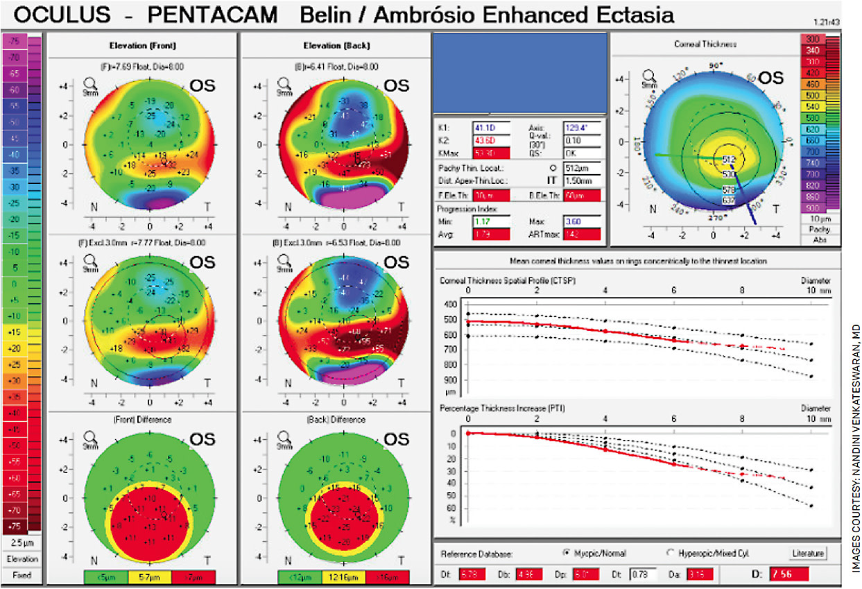
Indeed, tomography can be used to evaluate the anterior and posterior of the cornea, enabling specialists to better assess corneal astigmatism. It can also capture the earliest signs of keratoconus and corneal ectatic disorders before they manifest on topography, tomography’s two-dimensional imaging sibling (Figure 1).

“We know that forme fruste keratoconus is the greatest risk for developing postoperative ectasia, and there are several well-recognized signs on corneal tomography that can be used to screen those patients,” says Julie Schallhorn, MD, MS, a cornea, cataract, and refractive surgery specialist at UCSF Health, based at the University of San Francisco. Dr. Schallhorn also relies on her Oculus Pentacam tomographer to evaluate patient response to keratoconus treatment, particularly corneal crosslinking.
“Having all that data is important to see if a patient is a good candidate for any type of refractive surgery — not just those looking for LASIK, but also for cataract surgery patients who may want premium lenses,” agrees Kourtney Houser, MD, a corneal specialist at the Duke Eye Center in Durham, N.C. “If they need a corneal refractive touch-up after surgery, it’s important to have that data on the front end.”
In addition to the Pentacam, Dr. Houser uses the Galilei (Ziemer), which combines corneal tomography and topography in one unit.
“The Galilei not only uses tomography, but it also uses Placido disc imaging,” she says. “You can actually print off the Placido ring image or look at it on the machine or in your EMR, and use the rings reflected off the cornea to [assess] the quality of that image,” she says.
The Placido rings also help to diagnose ocular surface pathology; many specialists consider tomography to be an essential all-around corneal imaging modality.
Dr. Hura says that he conducted an informal survey of surgeons through the Refractive Surgery Alliance last year, “and 95% of respondents said they consider tomography critical for laser vision correction evaluations.”
OPTICAL COHERENCE TOMOGRAPHY
Optical coherence tomography (OCT), a variation of tomography once used only to assess the retina, is also available to image the anterior segment (AS-OCT). It can be used to evaluate the different layers of the cornea, the dimensions of the anterior chamber as well as evaluate corneal thickness, the shape of the stromal interface after lamellar corneal surgery, the association between host and corneal graft in keratoplasty, among other things.
“We’re using OCT to look at how deep corneal scars go into the depth of the stroma or to look at LASIK flaps in detail,” says Dr. Houser. “There are devices that’ll use the OCT for curvature data, but [I use it more] for qualitative images to get more angle data deeper into the eye and to look at masses and other pathologies.”
In addition to early signs of ectasia, Dr. Schallhorn finds AS-OCT useful for epithelial mapping to identify anterior basement membrane and other corneal dystrophies. “I use that routinely in my refractive surgery patients,” she says. Multiple OCT devices are currently on the market.
CORNEAL TOPOGRAPHY: AN OLDIE BUT GOODIE
Considering the wide-ranging capabilities of corneal tomography, it’s worth asking: why use the much older technology of corneal topography at all?
“It’s a good question, but corneal tomography doesn’t cover all the bases,” says Dr. Schallhorn.
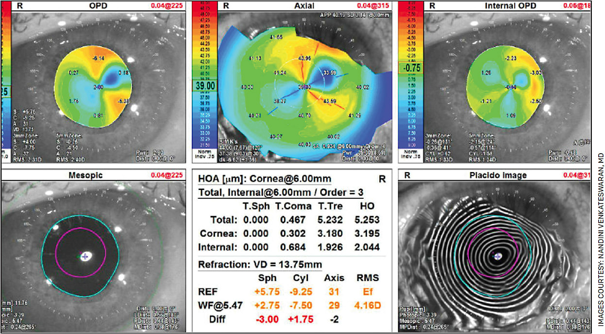
Placido disc topography fills multiple roles, including: screening, treatment and monitoring of corneal ectasia; preoperative IOL selection; post-keratoplasty astigmatism evaluation and management; and diagnosis of pterygia, corneal scars and Salzmann’s nodules (Figure 2). Citing the hypothetical case of a 50-year-old, -3-D patient who wants to be free of eyeglasses or contact lenses, Dr. Schallhorn says topography can help surgeons select from several interventions, including monovision IOLs, LASIK and refractive lens exchange.

“Looking at the internal aberrations with topography can be incredibly useful to make those determinations,” she says, adding that topography results can often help change patients’ minds about which intervention to pursue.
“I’ve had patients who are absolutely certain they want LASIK — until I show them the iTrace results,” says Dr. Schallhorn, whose imaging toolbox also includes a Tomey topographer.
Topography can also be used to obtain a second opinion on tomography, says Alanna Nattis, MD, a cornea, cataract and refractive surgeon at SightMD in Babylon, N.Y.
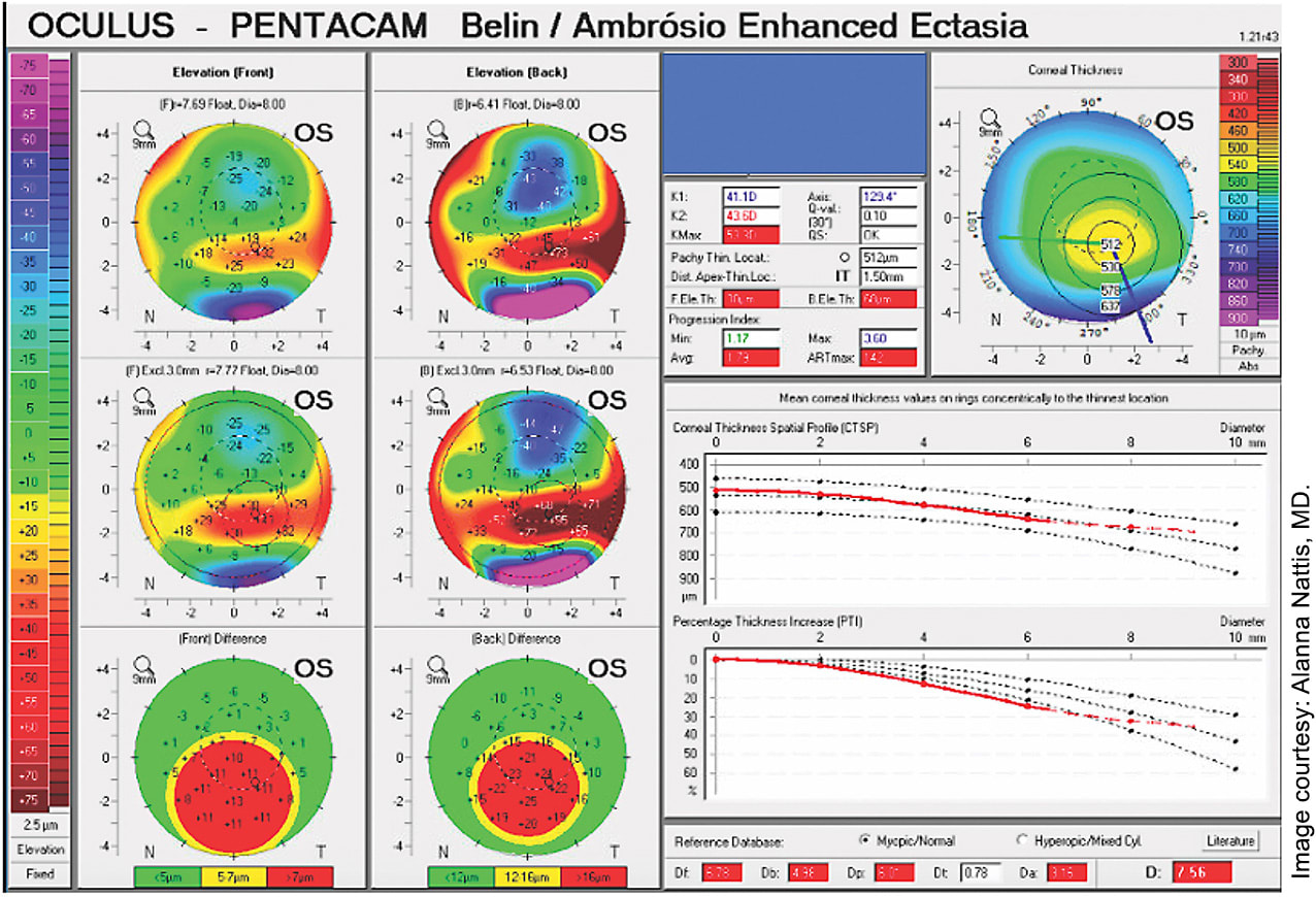
“When I’m doing a LASIK evaluation, I actually get a reading from the Pentacam and the Atlas (Zeiss) to see if one device picks up something that’s different or looks off,” says Dr. Nattis (Figure 3). She also employs the Orbscan (Bausch + Lomb) topographer, which she appreciates for its ability not just to take corneal images but also to evaluate their quality.

“Oftentimes you’ll get an off-center image, and it may look like you’re looking at an ectatic cornea when in fact it’s a normal cornea,” she says.
IF YOU CAN CHOOSE ONLY ONE …
Ultimately, given the unique capabilities of each modality, leveraging tomography and topography in practice can help provide a more comprehensive set of diagnostic imaging at a full-service ophthalmology practice. “As much as I would like to say that you can just go with one or the other, I don’t think that’s true, so I have both,” says Dr. Schallhorn.
All that said, it’s not always practical, or even necessary, for a practice to employ both technologies. For this reason, it’s important for practitioners to clearly define their goals. For example, ophthalmologists who plan to only perform cataract surgeries and not perform any laser vision correction or treat keratoconus may only need a topographer.
“But if you plan on doing everything, I’d say you need the topographer and the corneal tomography device,” Dr. Schallhorn says.
CORNEAL IMAGING KEEPS EVOLVING
If there’s any weakness to be said for corneal imaging in general, Dr. Hura says it’s the current dearth of “all-in-one” devices for tomography, topography and epithelial mapping. However, he is optimistic that these multi-modal diagnostic devices will emerge.
“Imaging devices continue to proliferate, and over the past couple of years at the American-European Congress of Ophthalmic Surgery meetings industry has indicated that they have been listening to surgeon feedback and are working on consolidating devices,” Dr. Hura says. “My hope is that in the not-too-distant future, surgeons won’t have to choose between what devices to invest in and that we will have more all-in-one options.” OM


