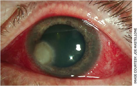
Our patient was a 49-year-old male with past medical history of insulin-dependent type 2 diabetes and hypertension and past ocular history of soft contact lens use. He presented with a one-week history of left eye pain that started when he was mowing the lawn.
He saw a local provider who initially diagnosed him with bacterial keratitis and started him on moxifloxacin 0.5% and tobramycin 0.3% alternating every hour. The patient reported worsening of left eye pain and redness despite compliance with ophthalmic antibiotic drops prescribed. Due to lack of clinical improvement, the patient presented in the emergency room one week after the inciting event, where he was cultured and started on fortified vancomycin 25 mg/mL and fortified tobramycin 14 mg/mL alternating every hour.
EXAMINATION
Examination of the patient showed the following:
- Distance VA with correction 20/25 OD, 20/200 PH NI OS
- Pupils: 3-2mm OU with no APD
- IOP (Tonopen, Reichert): 10 mm Hg OD, 11 mm Hg OS
Our patient’s slit lamp exam was notable for guarding left upper lid ptosis, 3+ conjunctival injection with no discharge, a 3.6-mm well-demarcated epithelial defect with a 4mm x 4mm round dense underlying infiltrate with feathery edges and no hypopyon (Figure). Corneal edema was noted with Descemet’s folds. No vitritis was noted on B scan.

Four days after re-culturing the patient and starting fortified antibiotics, preliminary microbiology results showed growth of “mold” (with identification to follow) and Bacillus species. The patient was started on amphotericin B 0.15% drops, and the aforementioned fortified topical antibiotics were continued. Fourteen days after initial culture, speciation results showed growth of Curvularia. Fortified antibiotics were stopped, and a generic fluoroquinolone was started to address the bacterial species. Natamycin 5% every hour was added to alternate with amphotericin B 0.15% every half-hour for treatment of Curvularia fungal keratitis. After starting natamycin 5%, the infiltrate decreased in size and the patient’s vision started to improve.
EARLY RECOGNITION IS KEY
This case delineates the diagnostic dilemma and challenges that can occur in the treatment of fungal keratitis, which is uncommon in the United States. The prevalence varies greatly with geographical locations but appears to have strong association in regions with warm humid climates.1 Clinical features of fungal keratitis can include feathery margins, satellite infiltrates and endothelial plaques; however, many cases do not present with these findings and are often misdiagnosed as bacterial keratitis.
It is widely noted that early recognition and treatment is critical in the management of fungal ulcers. One study further corroborates this, stating that a diagnostic delay greater than one week can extend the duration of treatment on average of 26 days. Additionally, fungal culture results may take a significant amount of time to complete; thus, it is critical to consider fungal keratitis in cases with lack of clinical improvement despite fortified antibiotic therapy or in cases of trauma with vegetable matter.2,3
WHERE CURVULARIA FITS IN
A recent systematic review published shows that the most common etiologies of fungal keratitis are Fusarium, Aspergillus, Curvularia and Candida species.4 Curvularia is classified as a dematiaceous mold most commonly found amongst soil and vegetable matter in warm, humid climates. Curvularia represents 4-9% of fungal isolates and is found in scrapings from patients with known fungal keratitis, making it the third most commonly recognized source of fungal keratitis.5-8 There are 40 species of recognized Curvularia with C. lunata and C. senegalesis representing more than 60% of all curvularia keratitis cases.9
Trauma involving soil or vegetable matter represents the most common source of curvularia infection. Other predisposing factors precipitating infection include the following: keratorefractive surgery, increased corneal exposure and, rarely, soft contact lens use. Additionally, Curvularia has been found to colonize <1% of human eyelids and 1-3% of healthy conjunctiva.10-12
Fungal keratitis from Curvularia has been shown to have a slower, more indolent clinical course with less inflammation than fungal keratitis from other sources. This slow clinical course often delays presentation and increases the risk for superinfection with concurrent bacterial colonization. One study illustrated that up to one-third of Curvularia cases were polymicrobial, as seen with our patient with concurrent bacillus species on culture.13
TREATMENT
In terms of treatment options, only one topical antifungal is commercially available in the United States: Natacyn (natamycin 5%, Santen). Others must be compounded. Natamycin is generally the preferred therapy against treatment of Curvularia species as it is best against filamentous species. Amphotericin B has also been shown to be efficacious in fungal keratitis but better for yeast species such as candida species.14
Stromal inflammation can be mild enough that the epithelial defect may resolve, making penetration of antifungals more difficult to achieve and requiring further debridement. There is also evidence that intrastromal and intracameral injection of antifungals such as voriconazole can be effective and well tolerated in recalcitrant fungal keratitis.15,16 Additionally, with larger infiltrates or fungal keratitis unresponsive to medical therapy, therapeutic penetrating keratoplasty must be considered. Care must be taken to minimize postoperative inflammation to prevent failure of the graft.17
CONCLUSION
Fungal keratitis is a serious complication of trauma with vegetable matter as seen in our patient. High suspicion for fungal etiology must remain on the differential when no improvement with antibiotics is seen for a presumed bacterial keratitis. Speciation also remains important for proper medical management. Curvularia is a less-common species in fungal keratitis and best responds to natamycin as seen in our patient. OM
REFERENCES
- Mahmoudi S, Masoomi A, Ahmadikia K, et al. Fungal keratitis: An overview of clinical and laboratory aspects. Mycoses. 2018;61:916-930.
- Wilhelmus KR, Jones DB. Curvularia keratitis. Trans Am Ophthalmol Soc. 2001;99:111-132.
- Khurana A, Chanda S, Bhagat P, Aggarwal S, Sharma M, Chauhan L. Clinical characteristics, predisposing factors, and treatment outcome of Curvularia keratitis. Indian J Ophthalmol. 2020;68(10):2088-2093.
- Ahmadikia K, Aghaei Gharehbolagh S, Fallah B, et al. Distribution, prevalence, and causative agents of fungal keratitis: A systematic review and meta-analysis (1990 to 2020). Front Cell Infect Microbiol. 2021;11:698780. Published 2021 Aug 26.
- Jan J-H, Ma DH-K, Tsai R-F. Keratomycosis: Ten years review. In: Shimizu K, ed. Current Aspects in Ophthalmology. Vol. 1. Proceedings of the XIII Congress of the Asia-Pacific Academy of Ophthalmology. Amsterdam, Netherlands:Excerpta Medica; 1992:339-342.
- Luque AG, Nanni R, de Bracalenti BJC. Mycotic keratitis caused by Curvularia lunata var aeria. Mycopathologia 1986;93:9-12
- Rosa RH, Miller D, Alfonso EC. The changing spectrum of fungal keratitis in south Florida. Ophthalmology 1994;101:1005-1013.
- Sandhu DK, Randhawa IS. Studies on the air-borne fungal spores in Amristar: Their role in keratomycosis. Mycopathologia 1979;68:47-52
- Roxman D, Jezernik K, Komel R. Ultrastructure and genotype characterization of the filamentous fungus Cochliobolus lunatus in comparison to the anamorphic strain Curvularia lunata. Microbiol Letters 1994;117:35-40.
- Ando N, Takatori K. Fungal flora of conjunctival sac. Am J Ophthalmol 1982;94:67-74.
- Sehgal SC, Dhawan S, Chhiber S, et al. Frequency and significance of fungal isolations from conjunctival sac and their role in ocular infections. Mycopathologia 1981;73:17-19. 108.
- Gupta AK, Dasgupta LR, Ramamurthy S, et al. Mycotic flora of normal conjunctiva. Orient Arch Ophthalmol 1972;10:128-131.
- Ahmadikia K, Aghaei Gharehbolagh S, Fallah B, et al. Distribution, Prevalence, and Causative Agents of Fungal Keratitis: A Systematic Review and Meta-Analysis (1990 to 2020). Front Cell Infect Microbiol. 2021;11:698780. Published 2021 Aug 26.
- Dorey SE, Ayliffe WH, Edrich C, Barrie D, Fison P. Fungal keratitis caused by Curvularia lunata, with successful medical treatment. Eye (Lond). 1997;11 (Pt 5):754-755.
- Garg P, Roy A, Roy S. Update on fungal keratitis. Curr Opin Ophthalmol. 2016;27:333-339.
- Kalaiselvi G, Narayana S, Krishnan T, Sengupta S. Intrastromal voriconazole for deep recalcitrant fungal keratitis: a case series. Br J Ophthalmol. 2015;99:195-198.
- Mundra J, Dhakal R, Mohamed A, et al. Outcomes of therapeutic penetrating keratoplasty in 198 eyes with fungal keratitis. Indian J Ophthalmol. 2019;67:1599-1605.


