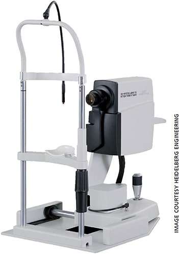
The Spectralis OCTA lets you comparatively track structure and function.
When OCT was introduced in the new millennium, it finally allowed ophthalmologists to obtain a cross sectional view of the retina. Only within the last five years have scan speeds become fast enough to detect a change in blood flow in the vessels, allowing for an en face view of perfused vasculature without the need for dye injection.
OCT angiography (OCTA), which has dramatically shifted the diagnosis and treatment paradigm of retinal vascular disease, is achieved by obtaining repeated consecutive B-scans at the same cross section. The motion contrast between static and dynamic tissues generates a complex signal that enables 3D visualization of the retinal and choroidal vasculature.1

THE NEW KID ON THE BLOCK
In September, Heidelberg Engineering announced that its Spectralis OCTA Module received FDA clearance, making it available here for new and existing upgradable diagnostic imaging devices.2 David M. Brown, MD, is a retina specialist at Retina Consultants of Houston, a practice that served as the principal investigator in the clinical trial for the FDA clearance of the technology. He reports that the OCTA Module has allowed him to comparatively track point-by-point references, from structural Spectralis OCTs gathered in his practice over the last seven years to new OCTAs taken with the OCTA Module. “This serial data analysis feature makes the Spectralis unique and indispensable.”
Heidelberg says the multimodal Spectralis offers the ability to combine OCTA with structural OCT, confocal scanning laser imaging and dye-based angiography in a single device.2 The 10° x 10° OCTA images have a lateral resolution of 5.7 µm/pixel to resolve the fine capillary network and an axial resolution of 3.9 µm/pixel for segmentation of the four histologically-validated retinal vascular plexuses.3 These include the nerve fiber layer plexus in addition to the superficial, intermediate and deep capillary plexuses.

CLINICAL UTILITY
“The superficial vascular complex is particularly useful for visualizing retinal vascular diseases such as diabetic retinopathy and retinal vein occlusion,” Dr. Brown says. “It allows you to easily see capillary dropout to assess why a patient’s acuity may be reduced and determine the visual prognosis.”
The deep vascular complex, which consists of the intermediate and deep capillary plexuses, is more finely tuned towards AMD. “Segmentation allows for viewing of the foveal avascular zone and macular capillaries that would otherwise be obscured by the superimposed layers with FA,” Dr. Brown says. “Although OCTA is improving logarithmically, the deeper layers are still prone to projection artifacts that make pathology harder to differentiate.” The Spectralis OCT offers TruTrack Active Eye Tracking, which prevents motion artifacts to ensure high quality images. Additionally, a projection artifact removal tool utilizes information from the superficial vascular plexus to remove artifacts via post processing for better views of the outer retina.4
“AMD, however, does not occur along a level plane,” explains Dr. Brown. “Drusen, inflammation and neovascularization cause bumps and changes among several planes or plexuses. Anti-VEGF therapy causes these planes to shift, making it even harder to differentiate the location of the pathology.” When the segments are confounded, the Spectralis OCTA provides an adaptive retinal slab feature — a continuous and interactive display of the retina, from the internal limiting membrane to Bruch’s membrane, for an all-encompassing view of the pathology.4
NEW DISCOVERIES
OCTA has also brought about the discovery of a new pathology, nonexudative choroidal neovascularization. The vascular contrast from flow within the new vessels was not seen prior to OCTA.5 In these cases, neovascularization occurs without leakage. According to Dr. Brown, “75% of these cases do not become exudative within a year, therefore anti-VEGF treatment may not be immediately required, and these patients can simply be monitored very closely using OCTA and structural OCT.”
CONCLUSION
Heidelberg Engineering Academy is available online to ensure ophthalmologists can confidently and efficiently use the technology.6 “Although it may take longer to acquire OCTA images with the Spectralis because of the required live eye tracking, the image quality and tracking provided by the structural OCT and functional OCTA have made this duo vital to the management of retinal vascular disease in my practice,” Dr. Brown explains. “As scan speeds continue to increase and software processing improves, OCTA will become an essential imaging modality.” OM
REFERENCES
- Jia Y, Bailey ST, Wilson DJ, et al. Quantitative optical coherence tomography angiography of choroidal neovascularization in age-related macular degeneration. Ophthalmology. 2014;121:1435-1444.
- Heidelberg Engineering OCT Angiography Module Now Available in the United States hhttps://www.heidelbergengineering.com/us/press-releases/heidelberg-engineering-oct-angiography-module-now-available-in-the-united-states/ . Accessed Nov. 10, 2018.
- OCT Angiography Module. https://business-lounge.heidelbergengineering.com/ca/en/products/spectralis/oct-angiography-module/?_ga=2.172436065.352044755.1541862681-1972351706.1493134963 . Accessed Nov. 11, 2018.
- Rocholz R, Teussink MM, Dolz-Marco R, et al. SPECTRALIS Optical Coherence Tomography Angiography (OCTA): Principles and Clinical Applications. 2018. Heidelberg Engineering GmbH.
- Lane M, Ferrara D, Louzada RN, et al. Diagnosis and Follow-Up of Nonexudative Choroidal Neovascularization with Multiple Optical Coherence Tomography Angiography Devices: A Case Report. OSLI Retina. 2016;47:778-781.
- Heidelberg Engineering Academy - SPECTRALIS OCT Angiography Module. https://academy.heidelbergengineering.com/course/view.php?id=292&_ga=2.24119928.352044755.1541862681-1972351706.1493134963 . Accessed Nov. 10, 2018.


