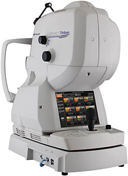
Device offers theoretical advantage in visualizing disease.
The FDA’s recent clearance of Topcon’s latest swept-source optical coherence tomography (SS-OCT) device for ophthalmic use offers clinicians new hope in screening and diagnostics, but how does it stack up to existing technologies?
“We have limited resources for managing patients, so when new technology comes out, we want to know, ‘Does it help the patient?’ and ‘Can we afford it?’” says Andrew Iwach, MD, clinical spokesman for the AAO.
Swept-source technology isn’t new, having been used in research devices for over a decade. But, Topcon’s DRI OCT Triton offers a few features that may improve outcomes in a commercially available unit.

FASTER SCANNING, LESS ROLL-OFF SENSITIVITY
The DRI OCT Triton boasts increased patient comfort, say users, but perhaps its most appealing feature is its faster scanning capability.
With an ability to scan 100,000 A-scans per second, Topcon’s device offers the fastest scanning available for commercial use, the company says. By design, such a feature should yield higher-quality images characterizing the tissues of the eye not visible to the naked eye.
However, at 15,000 A-scans above the most powerful spectral-domain OCT (SD-OCT) device previously available for use in the United States, whether the faster screening offers a clinical advantage that outweighs the cost remains unclear at this point, according to Jay Duker, MD, director of the New England Eye Center and professor and chairman of the Department of Ophthalmology at Tufts Medical Center and the Tufts University School of Medicine in Boston.
“The higher speed means you can cover more retina with your scan in the same amount of time. So, if you’re 15% faster, you can cover 15% more retina,” says Dr. Duker. “The more retina you cover, technically, the more disease you can pick up, although the truth is most of the disease we want to pick up is right in the center of the retina. So, it’s a theoretical advantage.”
The device utilizes an oversampling feature that eliminates noise by scanning tissue in the exact same position multiple times and editing out the variances. The end result is sharper imaging.
The DRI OCT Triton employs a swept-source laser for scanning. Unlike spectral-domain devices, swept-source devices use longer wavelengths, says Richard Spaide, MD, of Vitreous Retina Macular Consultants in New York. The 1050-nm waves can penetrate deeper into tissue than spectral-domain devices, which are currently the most widely used devices in the industry.
Swept-source devices show less roll-off in sensitivity with depth. This proves particularly helpful in visualizing through cataracts, Dr. Duker says, as the signal-to-noise ratio worsens with deeper spectral-domain scans, translating to poor image quality.1 Both Drs. Duker and Spaide noted that this feature helps elucidate the images of tissues better than the way it is characterized by spectral domain.
Dr. Duker says swept-source devices allow clinicians to capture images of the eye from the front of the retina to the back of the retina to the choroid without compromising sensitivity.
AS FOR THE PRICE TAG
Experts agree that the cost of new technology, when compared to the older SD-OCT technology, may cause practice owners to think twice.
“Any given instrument has a cost of goods that is very dependent on the number of units sold,” says Dr. Spaide. “A cheaper instrument would likely sell more than an expensive one; the more that are sold, the lower the cost to manufacture. So, there is a complex interplay between the cost, technology level and number of units sold.”
Moreover, very few swept-source devices are cleared for use in the United States, and the path to market here is a slower one than overseas. Other swept-source devices are used in the United States in research, including one that scans at speeds of up to 400,000 A-scans; but, these devices are not available for commercial use.
Topcon points out that as SS-OCT becomes more commonplace, prices will come down industry-wide.
LOOKING AHEAD
Technological advancements that improve screening and characterization of ocular tissue can benefit patients with early diagnosis of glaucoma, retinopathy, cataracts and other ocular complaints. Similarly, ocular abnormalities may enable clinicians to identify other non-ocular disease states that exhibit clinical manifestations that affect the eye. This could spawn greater interdisciplinary collaboration among health-care professionals through referrals, all thanks to enhanced visualization of ocular tissue.
“The eye is the only place in the body where you can see an artery or vein without an incision, so the eye may be an important biomarker for other diseases. We’re getting requests to collaborate with other specialties,” says Dr. Iwach.
Experts agree that OCT is an absolute necessity to modern-day clinical practice, and OCT devices such as the DRI OCT’s progeny will eventually dominate the market as the scanning speeds increase and the prices come down. “You can’t practice 21st-century ophthalmology without an OCT,” Dr. Duker says. OM
Relevant disclosures: Dr. Duker is a consultant to and receives research funding from Topcon. Dr. Spaide has received fees from Topcon.
REFERENCE
- Munk MR, Giannakaki-Zimmermann H; Berger L; Huf W; Ebneter A, et al. OCT-Angiography: a qualitative and quantitative comparison of 4 OCT-A devices. PLOS One. 2017 May 10;12(5):e0177059.


