Clearing payment hurdles
Experts offer three winning tips to help.
By Frieda Wiley, contributing editor
The ophthalmology community continues feeling the aftershock of payment challenges as health-care payer models remain in flux. Delayed or unremitted payment are becoming all too familiar, further exacerbated by increasing patient responsibility with the transition to high-deductible and high co-pay plans. Now, many ophthalmic practices are struggling to collect payments from patients — many of whom previously contributed minimal out-of-pocket payments to their health-care providers for services rendered.
“Originally, payment transactions occurred through B2B models where physicians billed the payer and were reimbursed directly by insurance companies. Patients rarely got involved,” says Mark E. Kropiewnicki, Esq., LLM, president and attorney at Health Care Law Associates in Plymouth Meeting, Pa.
To help ease payment collection woes, industry experts offer the following best practices.
1. Gain — and offer — clarity on each party’s expected payment prior to the patient’s appointment. When Carrie Jacobs, vice president of Chu Vision Institute in Bloomington, Minn., first noticed a surge in unpaid invoices in accounts receivable attributed to unmet patient deductibles and unpaid patient invoices, she and her team devised several strategies in efforts to curtail the growing trend.
“We revamped our policy to verify benefits on every patient and to collect payment at the point of service on the estimated out-of-pocket patient responsibility for all clinic visits and surgical procedures,” she says.
Ms. Jacobs also encourages practices to verify benefits and collect any responsible amounts due at the time of service to help eliminate surprises.
2. Train employees, and establish an inclusive protocol for collecting payment upfront. Along with employees in administrative roles, William Koch, COA, COE, CPC, administrative director at Texas Retina Associates, stresses the importance of training the entire staff, including physicians, on billing and payment collection processes. Mr. Kropiewnicki also recommends practices implement a working script for interactions with patients during financial transactions. Enacting protocols and collecting prompt payment reduce the risk for nonpayment, they add.
3. Recruit technology to facilitate payment collections. Accepting credit card payments increases the chances of collecting compensation for patients who may not have immediate funds available. Mr. Kropiewnicki points out that secure web payment options also increase the likelihood of collecting payment. He cautions that practitioners should note that employing such technology may deter certain groups of people — such as elderly patients and others who may have personal aversions to using technology to handle confidential information.
Collecting payment is never easy, but planning and preparation will help the process. OM
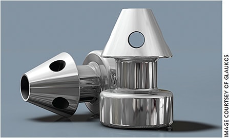
iStent inject approved by FDA
The Glaukos iStent inject received premarket approval from the FDA, based on results from its IDE pivotal trial. The iStent inject Trabecular Micro-Bypass System, designed to be used with cataract surgery to reduce IOP in open-angle glaucoma patients, achieved statistically significant reduction in unmedicated diurnal IOP in cataract surgery patients, according to Glaukos.
In the iStent inject cohort (387 patients), 75.8% achieved a 20% or greater reduction at 24 months, with a mean reduction of 7 mm Hg. Observed data showed that the iStent inject cohort achieved a 31% mean reduction in unmedicated IOP from an unmedicated mean baseline IOP of 24.8 mm Hg to 17.1 mm Hg. Also, the inject cohort’s overall rate of adverse events was similar to cataract surgery alone. Glaukos intends to start the iStent inject’s commercial launch in the third quarter. The iStent inject, which is already commercially available in other parts of the world, has been implanted more than 30,000 times. OM

Alcon’s Project 100 brings phaco to developing countries
The company will donate 100 Infiniti Vision Systems to Asia, Latin America and Africa in the next three years.
By Robert Stoneback, associate editor
Alcon will donate 100 of its Infiniti Vision Systems to clinics in developing areas of Asia, Africa and Central and South America. Donations of the Infiniti systems, used in phaco cataract surgeries, are part of the Alcon Cares program, Alcon Cares Project 100. The goal of Project 100 is to help underserved communities perform 200,000 phaco surgeries and train at least 400 doctors in phacoemulsification by the end of 2020, says Melissa Thompson, president of Alcon Cares and director of Corporate Social Responsibility.
Project 100 will begin this year in Asia before expanding to Central and South America in 2019 and then to Africa in 2020.
Alcon will work with regional partners in those locations to select clinics and hospitals for the donated Infiniti systems. Criteria for the facilities includes proficiency in phaco surgeries and adequate infrastructure to sustain an eye-care practice. Also, Alcon will partner with non-government organizations to train surgeons in the use of the Infiniti systems.
Alcon chose Asia as the starting point, says Ms. Thompson, due to the company’s “strong foundations for phaco surgeries” on that continent and relationship with non-government organizations to help identify and support local clinics.
Alcon expects to place about 30 units a year in the program and will determine allotment by patient need, demonstrated proficiency in phacoemulsification and doctor training.
“More than 90% of people with visual impairment and blindness reside in underserved, lower/middle income countries where health care access is limited,” says Ms. Thompson. “Whereas in developed countries cataract blindness represent 5% of eye diseases, it represents 50% or more in poor and/or remote regions. Further, eliminating blindness is one of the most cost-effective ways to fight poverty, as every dollar invested into prevention results in at least a four-dollar economic return.”
The Infiniti systems are “reprocessed” models, which were previously used in the marketplace and have been restored to original manufacturing specifications, says Ms. Thompson.
Previous donations from the Alcon Cares program include equipment to outfit surgical suites on the Orbis Flying Eye Hospital and Mercy Ships’ Africa Mercy. Between 2008 and 2017, Alcon Cares supported approximately 6,000 medical missions with $460 million in product donations. OM
LacryDiag marks Quantel’s entry into DED market
Non-contact device can perform four exams in four minutes.
By Robert Stoneback, associate editor
Quantel Medical has launched its LacryDiag diagnostic device, making it the company’s first entry into the dry eye market. An ocular surface analyzer approved by the FDA and compliant with the DED diagnosis recommendations in the DEWS II report, the LacryDiag can conduct four non-contact exams in four minutes. The exams, which together analyze the three layers of the tear film, are interferometry, non-invasive break-up time, tear meniscus and meibography of the upper and lower eye lids.
“With LacryDiag, ophthalmologists are now able to diagnose in a few minutes the three tear film layers to select a personalized treatment for the patient,” said Jean-Marc Gendre, CEO of Quantel Medical, via press release.
The LacryDiag can identify the presence of DED as well as whether it is evaporative or aqueous deficient DED, according to Delphine Southon, dry eye product manager for Quantel Medical. The LacryDiag includes a yellow filter for staining to detect damage to the cornea, conjunctiva and eyelids, a feature not found on other models of dry eye analyzers, according to Ms. Southon. OM
CenterVue announced that the company received FDA approval for its Eidon FA, which adds fluorescein angiography capabilities to the CenterVue Eidon platform. The fully automated system includes multiple imaging modalities. The Eidon FA features confocal technology, which allows for high-resolution retina images (15 μm) to detect and follow retinal blood flow. Also, its 60° widefield images allow for high quality visualization of the retinal midperiphery.


