INNOVATION
A Streamlined System
How Marco technology helped to improve outcomes and efficiency in my practice
BY MARIA C. SCOTT, MD
More than 20 years ago, I remember walking into my first exhibit hall during the annual meeting of the American Academy of Ophthalmology and being mesmerized by all of the gadgets, instruments, and equipment. Even today, I’m overwhelmed by the vast array of technology available for providing superior ophthalmic care. I tend to be frugal in my personal purchases, but when it comes to ophthalmic equipment and the promise for better patient outcomes and improved efficiencies, all restraint goes away. I think many of us share the same philosophy — ophthalmologists love “toys.” But today, with declining reimbursement and space constraints, I must be more selective in my purchases, using a critical eye when promised improved outcomes and faster throughput.
More Accuracy, Less Space
In 1996, I bought a Marco Epic Refraction System after being introduced to it by architect Jeff Eckert, who described how he maximized space using the Epic. With a 3’6” × 5’6” footprint, the size of the refracting lane is reduced significantly. In fact, the Epic can fit comfortably into a 6’ × 8’ space.

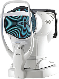
OPD-Scan III
In addition to the convenient size, I loved offering patients a high-tech experience that delivered a “wow” factor. I purchased my second Epic about 3 years later. It’s hard to believe that we still use these machines every day, nearly 20 years later.
Besides the Epic machines, we later purchased two OPD-Scan III machines from Marco. We use these on all cataract evaluation patients. With our OPD scanners placed next to our LenStars (Haag-Streit) and IOLMaster (Zeiss), we can gather auto refraction, auto keratometry, corneal topography, wavefront analysis, pupillometry, and placido disc photo-graphy in 10 seconds per eye with just one machine. It also saves time by eliminating trips to multiple testing instruments. Besides the speed, this approach also improves accuracy because there’s less drying of the eye surface.
Our Process
In our practice, we perform the OPD testing before any other testing. The cataract technician takes three scans and compares AKs to Lenstar and IOLM keratometry. If there’s a discrepancy, it usually indicates ocular surface disease of some kind. This can be confirmed with placido disc images, which can also be used to illustrate the problem to the patient.
With detailed corneal analysis, I can quickly assess corneal astigmatism, separate anterior corneal from internal astigmatism, locate the axis of astigmatism, and determine whether the astigmatism is regular or irregular. Although it cannot calculate posterior and lenticular cylinder separately, it can mathematically help you determine the difference by selecting “subtract prism” in the sub-menu on the tools bar. Again, sharing the astigmatism graphic with patients makes it easy to explain what astigmatism is, the different places it can occur, and how it can be treated.
Calculating angle kappa and corneal higher order aberrations (HOA) helps to determine which patients may not be good candidates for multifocal lens implants. Spherical aberration greater than .32 or angle kappa greater than .42 for most multifocal lenses may predict poor vision quality after multifocal IOL implantation. If the patient has a large angle kappa, the center of the pupil is no longer the point through which a fovea-centric ray of light passes. The patient will see through the rings of the IOL off to the side, which would increase complaints of glare and halos.
Post-LASIK patients who had myopic treatments before custom LASIK or high myopic treatments usually have an HOA well above .32 with some as high as >1.0. These patients would most likely be poor multifocal candidates. Selecting an IOL that neutralizes the eyes’ spherical aberration would help to improve image quality. I no longer assume multifocal IOLs aren’t a good fit for post-LASIK patients. Instead, I make an informed decision based on the patient’s OPD results.
WHEN THE OPD-SCAN III IS COMBINED WITH THE TRS 5100 AUTOMATED REFRACTION SYSTEM, TECHNICIANS CAN QUICKLY PERFORM A WAVEFRONT-OPTIMIZED REFRACTION, SAVING 3 TO 5 MINUTES PER EXAM. THEY CAN ALSO SHOW PATIENTS THE DIFFERENCE BETWEEN THEIR SIGHT WITH THEIR OLD EYEGLASSES AND THE NEW PRESCRIPTION.
Post-LASIK patients also pose a challenge in determining the correct IOL power. Typical post-myopic treatments will yield a hyperopic surprise if an adjustment in IOL selection is not made. In addition, some patients are unsure whether they were hyperopic or myopic before LASIK surgery. Looking at the topography and the induced HOAs may give us a clue. The OPD-Scan III calculates the average pupil power, which can be used in formulas to calculate the “new” IOL power.
The pupillometer shows photopic and mesopic pupil size giving day/night differences in prescriptions. The day/night summary can tell us which patients have nighttime driving problems, which may necessitate a separate nighttime prescription. Some IOL choices are pupil dependent, particularly multifocal lenses. The pupillometer can help make the best lens choice for the patient.
The retro-illumination feature can document and illustrate the existence of cataracts and can be used to evaluate the placement of a toric IOL postoperatively.
Point Spread Function (PSF) can differentiate whether distortion is corneal or lenticular. Patients with substantial corneal distortion can be educated preoperatively to be prepared for possible residual ghosting even after the cataract is removed.
Patient Education
Preoperative patient education is key to postoperative patient satisfaction. The OPD-Scan III provides a large quantity of diagnostic information in one setting and helps customize the IOL to each patient. Through the OPD-Scan III viewer station software or integration with EMR or image management systems, it is possible to show patients and family members maps while in the exam room to help them understand the disease process and accept your recommendations. The OPD-Scan III also documents preexisting conditions.
When the OPD-Scan III is combined with the TRS 5100 automated refraction system, technicians can quickly perform a wavefront-optimized refraction, saving 3 to 5 minutes per exam. Using the ETDRS chart or sailboat photograph, they can also show patients the difference between their sight with their old eyeglasses and the new prescription. This can also be used to illustrate that new glasses may not improve vision if a cataract is present. The refraction data populates into appropriate fields in the electronic health record, avoiding the risk of entry errors and saving time. The determination of cylinder and axis using prismatic split-screens is the quickest method used. The system offers testing for near point of accommodation, stereopsis, convergence, phorias, and tropias.
A Real Game-changer
In summary, I think the multi-modality Marco automated refraction equipment passes the sniff test for efficiency, increased throughput, improved outcomes, and multipurpose utility. The data is presented to both the surgeon and the patient in a way that supports and speeds clinical decision-making. It provides a complete assessment of the optical pathway, making higher-level problem solving possible with better patient flow and efficiency, while optimizing surgical outcomes. ■
Maria C. Scott, MD, is founder and medical director of Chesapeake Eye Care and Laser Center and medical director of TLC Laser Eye Center in Annapolis, Md. She is a member of the OOSS Board of Directors. |


