Quick Hits
Xiidra (Lifitegrast) wins FDA approval
The dry-eye prescription-therapy drought has ended.
By René Luthe, Senior Editor
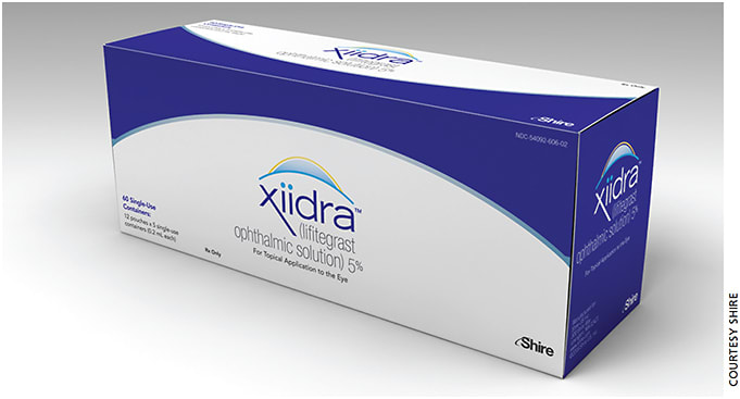
Dry eye care’s long dry spell for new prescription treatments ended in early July when the FDA approved lifitegrast ophthalmic solution 5% (Shire). It’s the first such approval since Restasis (cyclosporine ophthalmic emulsion 0.05%, Allergan) entered the U.S. market in 2002.
Lifitegrast — under the brand name Xiidra — also has the distinction of being the only prescription eyedrop indicated for the treatment of both signs and symptoms of dry eye disease (DED) in adult patients. The novel small molecule integrin antagonist works by inhibiting T cell-mediated inflammation by blocking the binding of two critical cell surface proteins (lymphocyte function-associated antigen 1 and intercellular adhesion molecule 1); the result is an overall reduction in inflammatory responses.
Xiidra will be marketed in individual-unit doses for its b.i.d. application. The ampule is similar to some of the commonly used artificial tears, notes Shire’s Robert Dempsey, vice president, eye care. And it’s preservative-free, an important selling point for dry eye sufferers. “We do not want to have preservatives applied to the ocular surface of patients who present with dry eye,” Mr. Dempsey says.

Gearing up for “tremendous opportunity”
Shire expects to launch Xiidra in the third quarter of this year.
According to Mr. Dempsey, that launch will include a direct-to-consumer campaign to educate patients on the condition of dry eye and how it affects their lives. He estimates that 12.5 million people in the US have diagnosed symptoms associated with DED, and of that group, 1 million to 1.4 million have been or are being treated with the only other prescription product available here — cyclosporine, while the rest are being treated with over-the-counter artificial tears. “So there is tremendous opportunity to treat these symptoms associated with DED. We’ve done an extensive amount of research with consumers as well as eye-care professionals, and it really is the symptoms of DED that are driving patients to the doctor’s office,” Mr. Dempsey says.
Along with training and deploying its sales organization, Shire will be engaging with health insurance companies over the next few weeks to begin the process of gaining access to their formularies once the product is available.
High hopes
Two eye care veterans in the struggle to identify and treat DED voiced their appreciation for a new prescription-strength weapon. “Lifitegrast represents a novel approach to address ocular surface inflammation, which is a core mechanism of dry eye,” says Frank W. Bowden, III, MD, of Bowden Eye & Associates, Jacksonville, Fla.“We are anxious to see how it will complement existing dry eye therapeutics.”
The therapy shows an important advantage, at least so far, according to Marguerite B. McDonald, MD, of Ophthalmic Consultants of Long Island, N.Y. “There is no evidence [within the time frame of the studies] that Xiidra’s effect wears off with time,” she says. And Xiidra may yet offer other ocular benefits. “There may indeed be other ophthalmic uses now that post-approval studies can commence with IRB [institutional review board] approval for other indications. I am very happy that Xiidra was approved; we need all the ammo we can get in the battle against dry eye.”
And that ain’t all
Though the long slog in quest of Xiidra’s FDA approval is over, the company expects to continue seeing a lot of the regulatory agency: Shire has four other programs in development. A treatment for infectious conjunctivitis is now entering Phase 3 trials, while another, while not meeting its primary endpoint to reduce the severity of retinopathy of prematurity, will be the subject of a near-term meeting with the FDA regarding the trial’s success in meeting its secondary endpoints related to severe bronchopulmonary dysplasia and severe intraventricular hemorrhage. A third, early-stage program addresses glaucoma. “We have yet to classify the MOA, but it has shown tremendous signals in animal models,” says Mr. Dempsey.
Shire’s fourth program, which the FDA has given fast-track status, focuses on autosomal dominant retinitis pigmentosa.
“We are building a comprehensive franchise for the anterior segment, the posterior segment and inherited retinal diseases,” says Mr. Dempsey. “It’s an exciting time for the organization to have an anchor program like Xiidra enter the market, and we’re building up the commercial, medical and R&D capabilities.” OM
Is race an outdated way of looking for disease?
A recent study suggests a more personalized look at a patient’s history.
By Robert Stoneback, Associate Editor
Scientists and physicians have used race for years as a way to measure risk for genetic diseases, but research argues that such a broad category might be detrimental for patients.
The paper, from Science, does not dispute that people of different races face different health risks. However, to view those health problems purely “as an outcome of biology” does not reflect what is known about human genetics, according to one of the researchers, Dr. Michael Yudell, PhD, associate professor and chair of community health and prevention at Drexel University.
Dr. Yudell and his co-authors warn that a reliance on race to detect diseases can lead physicians to misdiagnose some ailments. For instance, sickle cell anemia is considered a “black disease,” but it may go unnoticed when it appears in people of other races, as physicians are not expecting to find it, Dr. Yudell continues.
Race as a starting point
Nathan Radcliffe, MD, clinical assistant professor of ophthalmology NYU Langone Medical Center, says that race can be an important starting point for diagnosing glaucoma’s risk factors, but physicians should then move beyond that to understand the driving risk factors for a patient.
While Dr. Radcliffe’s research shows that corneal thickness differs between blacks, whites and Hispanics, level of thickness is often a better indicator of glaucoma than a person’s race.
“If you fully account for corneal thickness, then race no longer matters, and I think that’s where we want to get, to understand our risk factors well enough,” he says.
His 2012 paper published in Acta Ophthalmologica examined corneal hysteresis (CH) and central corneal thickness (CCT) in black, whites and Hispanics. While there were differences between the races — with blacks shown to have lower levels of CH and CCT on average compared to whites and Hispanics — there was also significant variation between CH within the racial groups. This suggests that CH may be preferable for evaluating glaucoma risk and other corneal properties within these groups.
If a physician lacks access to tools such as corneal thickness, CH levels or family history, then race can be used as “a starting point, and that may give you some justification for pushing ahead for other diagnostic tests,” Dr. Radcliffe says.
Ancestry & precision medicine
Rather than use race, Dr. Yudell and his colleagues recommend using a patient’s ancestry to judge their risk for disease.
Again using sickle cell anemia as an example, it would be more accurately described as a disease found frequently in ancestry groups from certain parts of Africa, the Middle East and the Mediterranean, according to Dr. Yudell.
The 2016 Science article findings suggest that “precision medicine,” also called individualized genomics, could be the future of treating diseases, according to Robert DeSalle, PhD, one of the geneticists who helped author the paper. Dr. DeSalle serves as a professor at the American Museum of Natural History’s Richard Gilder Graduate School and curator of the museum’s molecular schematics. Precision medicine, which is designed to tailor healthcare to individual patients, is already being investigated as possible treatments for AMD and leber congenital amaurosis, according to a paper published in the March issue of Investigative Ophthalmology and Visual Science. Predictive tests still need to be developed so that precision medicine can be tailored to individual needs, according to the study.
“The most important thing we’re talking about in this paper is that race is not a good proxy for understanding an individual’s genes and how they might relate to health outcomes,” Dr. Yudell says. “When we diagnose by using race, we are diagnosing based on a probability.” OM
Coming to U.S. presbyopes: The Raindrop inlay
With Raindrop’s recent FDA approval, surgeons now have two options for near-vision correction.
By René Luthe, Senior Editor
The nation’s approximately 111 million presbyopes1 now have a new option to replace those dreaded “readers”: on June 29 the FDA approved ReVision Optics’ Raindrop Near Vision inlay.
According to the FDA’s indication, the Raindrop is applicable for patients between 41 to 65 years of age with emmetropic eyes who cannot focus clearly on near objects or small print. These patients need reading glasses with +1.50 to +2.50 diopters of power, but do not need glasses or contacts for clear distance vision. The data ReVision Optics submitted apparently impressed regulators. The company, which received approval eight months after submitting its PMA, will not need advisory panel review. It demonstrated that the inlay was safe and effective based on a clinical trial of 373 patients. After two years, 92% of the patients included in the final analysis (336 out of 364) had 20/40 vision or better at near distances. Adverse events included epithelial ingrowth in less than 3% of the eyes, inflammation in 1.6%, the exchange of 19 inlays and the report of glare and halos from three patients — the latter being fewer than with LASIK, says Mike Crompton, ReVision Optics’ vice president, regulatory affairs and quality/chief compliance officer.
Douglas D. Koch, MD, a lead medical monitor for the FDA trials, calls the Raindrop “a superb option for treating presbyopic patients. The procedure is remarkably easy to learn — well within the scope of any LASIK surgeon. I felt comfortable with it after two cases.” An added bonus, he says, is that surgeons won’t need to purchase special testing equipment.
“Follow-up is important to monitor healing and ensure that patients adhere to the regimen of postoperative corticosteroid drops.”
Region of power
The tiny device — 2.0 mm diameter and approximately 30 microns thick — resembles a contact lens. It is made of a proprietary hydrogel material with a refractive index similar to that of the cornea. After making a femtosecond laser-created flap into the cornea of the patient’s non-dominant eye, the surgeon inserts the device, implanting it onto the stromal bed of the cornea. The surgeon then centers the Raindrop over the light-constricted pupil, thereby creating a prolate-shaped cornea.
This reshaping provides a region of increased power for focusing on near objects, resulting in improvement in near vision. Because it is transparent, it does not restrict the amount of light that reaches the retina. The Raindrop is self-contained: the device is preloaded into its inserter, bypassing the need for additional capital equipment (outside of the femtosecond laser).
The procedure takes approximately 10 minutes, says ReVision.
Bad matches, major differences
As with any device, some patients are not good candidates for the Raindrop; the FDA says contraindications include dry eye, corneal disease such as keratoconus, certain autoimmune diseases, insufficient corneal thickness to withstand the procedure, uncontrolled glaucoma or diabetes, a herpes eye infection or pregnancy.
As for how the Raindrop compares to the only other FDA-approved implant for the correction of presbyopia, AcuFocus’ KAMRA inlay, surgeon Martin L. Fox, MD, notes important distinctions.
“The Raindrop and KAMRA differ considerably in their characteristics, mechanism of action and recommended implantation techniques,” he says. Implantation of the Raindrop requires placement of the inlay over the pupil, “under a LASIK flap that needs to be at least one third of the corneal thickness, or 150 to 160 microns,” Dr. Fox points out. “KAMRA surgery, on the other hand, consists of inlay placement in a narrow corneal pocket created by a femtosecond laser at 40% corneal pachymetry,” making it possibly a better choice for LASIK patients who have thin corneas, he says.
Gearing up for the U.S. market
ReVision Optics says it is expanding its sales force and is planning to introduce Raindrop to U.S. ophthalmologists in the third quarter of this year. Surgical training will include a wet lab and an online training module, says company CEO and president John T. Kilcoyne. He doesn’t rule out direct-to-consumer advertising in the future.
Also in the future: pseudophakic eyes could be the Raindrop’s next market. According to Mr. Crompton, outside the United States the Raindrop has already won regulatory authorization for patients who have had cataract surgery. These jurisdictions include the European Union and Australia. “We are working on gathering data for the FDA,” with an IDE clinical trial on pseudophakic eyes, he says. OM
REFERENCE
Size does matter
With 27-gauge vitrectomy, surgeons can take a less invasive approach.
By Louise Gagnon
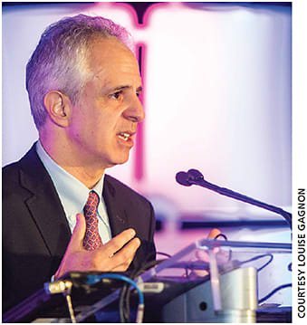
Describing the evolution in microincision vitrectomy surgery at the annual meeting of the Canadian Ophthalmological Society, Carl Regillo, MD said 27-gauge vitrectomy is almost always a sutureless procedure in contemporary vitreoretinal surgery, and it offers ophthalmologists the opportunity for straight rather than angled entry.
Dr. Regillo, director of retina service at Wills Eye Hospital and professor of ophthalmology at Thomas Jefferson University in Philadelphia, Pa., said the push to smaller incisional approaches in ophthalmology is consistent with less invasive approaches in medicine. “These approaches usually translate to faster recovery [for patients],” he said.
Some concerns about a sutureless approach leading to increased rates of endophthalmitis have been allayed, said Dr. Regillo. “The way things have evolved, there is maximum wound integrity through refining of surgical techniques, which have kept endophthalmitis rates very low.”
The 27-gauge instrument allows convenience of entry that the larger instruments do not, he said.
“With the 27-gauge, you can go in straight and not have to worry about an increase in the rates of leaks,” said Dr. Regillo. “If you can go straight in, it affords an easier approach to the tissues. With the 23- and 25-gauge [instruments], to make the wound secure and not leak, [entry] has to be angled. If you go straight in, you will get excessive leaks and excessive post-operative complications, including infection.”
Studies, both in vitro and in vivo, definitively show that the entry point when using 23- and 25-gauge vitrectomy has to be angled, said Dr. Regillo.
The original 20-gauge instrument possesses a 0.9-mm diameter, creates a significantly bigger wound, results in disruption of the conjunctiva and requires suture infusion. In contrast, the development of 23-gauge and 25-gauge instrumentation led to smaller surgical wounds with less tissue disruption, noted Dr. Regillo.

Carl Regillo, MD
A study that compared 25-gauge transconjunctival sutureless vitrectomy (TSV) and 20-gauge pars plana vitrectomy demonstrated, using ultrasound biomicroscopy, that the 25-gauge TSV produced a faster decrease of surgically-induced keratometric astigmatism related to rapid cicatrization of sclerotomy sites, according to a 2010 Cornea article by Avitabile et al.
A disadvantage of the 27-gauge instrument is its pliability, coupled with slower gel removal during a vitrectomy. “There is a learning curve in trying to work around the flexibility [of the 27-gauge],” Dr. Regillo said. “It is something that surgeons need to be aware of. It is undesirable [if they are too flexible]. You want your instruments to be stiff, so they move precisely where you need them to go.”
The development of smaller gauge instrumentation can be attributed to advancements in lighting and wide-field viewing systems, said Dr. Regillo. At present, the 27-gauge vitrectomy does not represent the standard vitrectomy approach, he said. “Retinal specialists are currently using the 23-gauge or 25-gauge platform as a primary platform,” he said. “They both perform very well and offer the advantages of sutureless surgery and faster healing.”
More challenging clinical cases, such as proliferative diabetic retinopathy, are a good fit for the 27-gauge platform. “Where it offers a significant advantage is complex tractional (retinal) detachment scenarios,” he said. Dr. Regillo estimated that the average vitreoretinal surgeon deals with a complicated tractional retinal detachment a few times per month, and that number can be higher or lower depending on the number of diabetic patients in a given practice.
However, Dr. Regillo did recommend using the 23- or 25-gauge platforms for macular holes because he does not see the advantage of using 27-gauge for that clinical scenario.
Asked about making the transition to the 27-gauge instrument, Dr. Regillo said it is easier to start as a 25-gauge instrument user.
In addition, Dr. Regillo said clinicians who use a contact, widefield viewing system will likely find it to be a simpler transition. “Surgeons who use that technique are more likely to be able to transition (to the 27-gauge instrument],” he told fellow clinicians. “I am a non-contact, widefield [viewing system] user, so I rely a lot on torquing the globe to see the peripheral retina. That is when you will get some [instrument] bending, and you might not like the 27-gauge [instrument] initially.” OM
Take the stepladder approach to uveitis
C. Stephen Foster, MD, on the latest strategies to treat this condition.
By Laird Harrison, Contributing Editor
Corticosteroids can provide good short-term relief from the inflammation caused by uveitis, but when used long-term they can cause serious side effects to most of the body’s major organ systems, including the endocrine and cardiovascular systems.1
A stepladder approach that starts with the least aggressive treatments can cure many uveitis cases without long-term reliance on corticosteroids, says C. Stephen Foster, MD, president and CEO of the Massachusetts Eye Research & Surgery Institution in Waltham, Mass.
“The main point to the general ophthalmologist community is to stop doing what people have been doing for the past 70 years. Namely, treating people with uveitis only with corticosteroids. The uveitis specialty community stepped away from that model of care about 30 years ago.”
The FDA’s approval of Humira (adalimumab, AbbVie) on June 30 to treat non-infectious intermediate, posterior and panuveitis highlighted the availability of alternatives to corticosteroids.
Tecnis Symfony IOL receives FDA approval

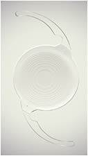
The FDA approved Abbott’s Tecnis Symfony IOL, the first extended depth of focus lens in the US. The Symfony is designed to provide continuous, full-range, quality vision following cataract surgery while helping presbyopic patients focus on near objects, Abbott says. The FDA’s approval also includes the Tecnis Symfony Toric IOL for patients with astigmatism.
The need is growing
Uveitis is the third-leading cause of worldwide blindness, yet few physicians specialize in treating this condition, says Dr. Foster, who is on the board of directors of the American Uveitis Society.
“There might be 100 appropriately trained people interested in uveitis in the United States,” Dr. Foster says.
And there’s a monetary motivation. In one recent study, annual medical costs for people with non-infectious intermediate, posterior or panuveitis totaled $7,790, of which $5,975 was spent for outpatient services. Annual pharmacy costs totaled $5,151.
For a matched sample of patients without uveitis, annual medical costs were $2,645, of which $1,997 was spent on outpatient services.2
Start at the bottom rung
Successful treatment of uveitis often depends on successful diagnosis of the etiology. In another recent study, researchers at the University of Virginia, Charlottesville, found that the HLA-B27 gene accounts for much of the morbidity associated with uveitis, and many patients with this gene present with systemic signs and symptoms.3 Other autoimmune diseases, however, may also be diagnosed with laboratory testing.
These researchers found that the most cost-effective initial diagnostic evaluation for Medicare patients was rheumatoid arthritis, followed by syphilis, Bartonella, Granulomatosis with Polyangiitis and polyarteritis nodosa. For Medicaid patients, HLA-A29 was the most effective initial diagnostic evaluation followed by HLA-B27, rheumatoid arthritis and toxoplasmosis.
General ophthalmologists and retina specialists typically default to using corticosteroids because they don’t know how to use the more sophisticated treatments that professional guidelines call for, he says.
Dr. Foster stressed the importance of starting with the least aggressive therapy for a specific uveitis etiology and severity and advancing to stronger treatments as needed. The lowest rung on the ladder consists of nonsteroidal anti-inflammatory drugs (NSAIDs), followed by classic immunomodulatory therapy, including corticosteroids. Above that, Dr. Foster lists peripheral retinal cryopexy or laser with pars planitis, then biologics and cytotoxic drugs. At the top sits pars plana vitrectomy. Topical, regional and systemic corticosteroids remain the mainstay of treatment, Dr. Foster says. Where appropriate, these can often be combined with cycloplegics and mydriatics. Clinicians should monitor patients closely and escalate treatment when a regimen fails to control inflammation, either by switching to a different drug or adding one, he says.
These treatments have allowed clinicians to cure many forms of uveitis defined as a sustainable remission with eventual withdrawal of all immunomodulatory medication and no relapse of ocular inflammation for five or more years, according to Dr. Foster.
Dr. Foster and his colleagues have achieved this sort of remission by using one or more immunomodulatory agents without tapering any goal-achieving drugs two to three years, and tapering slowly after that point.
The remission should occur without any steroids because they could mask any residual inflammation that might indicate a failure of the immunomodulation, he says.
Clinicians, he suggests, should familiarize themselves with NSAIDs, corticosteroids, mydriatic and cycloplegic agents, antimetabolites, calcineurin inhibitors, biologic response modifiers, alkylating agents and combinations of these drug classes, since all may play a role in uveitis treatment.
Conclusion
Surgery may help patients who don’t improve with medical treatment. Most uveitis patients, though, will recover well with the appropriate medical treatment, he says. “All of these things can be used for years without high risk of side effects. The rheumatologists have known this for many years, and their use of corticosteroids plummeted many years ago. Their mantra became ‘the mission is remission,’” he says, adding that physicians treating uveitis should not settle for less. OM
REFERENCES
1. Oray M, Abu Samra K, Ebrahimiadib N, Meese H, Foster CS. Long-term side effects of glucocorticoids. Expert Opin Drug Saf. 2016;15:457-465.
2. Thorne JE, Skup M, Tundia N, et al. Direct and indirect resource use, healthcare costs and work force absence in patients with non-infectious intermediate, posterior or panuveitis. Acta Ophthalmol. 2016 Mar 2. doi: 10.1111/aos.12987. [Epub ahead of print]
3. Gupta SK, Bajwa A, Wanchek T, Reddy AK. Cost-effectiveness of laboratory testing for uveitis. Austin J Clin Ophthalmol. 2015;2:1053.


