SPOTLIGHT ON TECHNOLOGY & TECHNIQUE
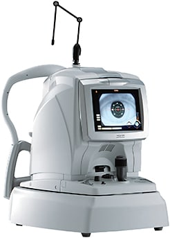
OCT device lets users capture precise retina, choroid images
With Nidek’s RS-3000 Advance, “You have the scan on the screen and away you go.”
By Zack Tertel, Senior Associate Editor
James S. Lewis, MD, an anterior segment specialist in Elkins Park, Pa., says he has experience with multiple OCT devices.
But since purchasing Nidek’s RS-3000 Advance OCT system, designed for evaluation of the retina and choroid, Dr. Lewis says his other devices are “collecting dust.”
The RS-3000 Advance provides an attention to detail that allows users to make proper diagnoses while other functions help increase office efficiency, Dr. Lewis says. “I can see the layers of the cornea, angle, choroid and retina in incredible clarity, and it’s extremely simple to use.”

PROCEDURE
Patients are positioned on the device’s single chin rest. Then they look straight ahead, which makes the procedure easy for patients, says Darrell Genstler, MD, of Genstler Eye Center in Albany, Ore.
“Most of our patients are older, so positioning them and getting them in and out is always an issue,” he says.
Once in place, patients follow large images, such as plus signs, that operators can move or change the color of to keep patients looking in the right direction. Also, the RS-3000 Advance features its Torsion Eye Tracer technology. This assists in capturing accurate images even when patients move, says Dr. Lewis.
“It doesn’t just notify you that you’re off target — it actively re-centers you. Then, it continues acquiring clear images.”
FOLLOW-UP
After the procedure, physicians can analyze and compare images via the RS-3000 Advance’s NAVIS-EX image filing software.
Dr. Lewis says he appreciates that this feature allows him to sit with the patient in the exam room and view the images without tying up the room or the device. This level of seamless connectivity allows physicians to analyze and compare images on any computer in the office.
“I can pull up images, manipulate them and counsel the patient in a relaxed a private setting,” says Dr. Lewis.
With this method of image review combined with its scan quality and operation system, the RS-3000 Advance has allowed increased patient flow and precise diagnoses, say those interviewed.
“It takes very little time, dilated or undilated,” Dr. Genstler says. “You take the patient in there, you get the patient back in the room, you have the scans on the screen and away you go.” OM


