3-D in the OR: a better view from many angles
Early adopters are finding advantages ergonomically, surgically and educationally.
By Karen Blum
| About the Author | |
|---|---|
|
Karen Blum is a medical writer based in Owings Mills, Md., who specializes in eye care. |
|
While the concept of three-dimensional teaching in ophthalmic surgery has been bandied about for two decades, the first systems to hit the marketplace a few years back weren’t quite perfect. They had some lag time between the surgeon’s view through the microscope and the image projected on a monitor, or required the purchase of system-specific monitors or other equipment that may have put some customers off.
But now, with faster processors and other improvements in technology, and the availability of 3-D televisions at local electronics stores, some eye surgeons who have recently taken the plunge say they’re finding the systems to be indispensable.

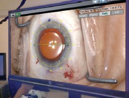
3D video can show details, such as IOL positioning, in high definition, as seen here in the OR of Jacob Moore, MD.
“It’s the world’s greatest teaching tool,” says Richard Mackool, MD, director of the Mackool Eye Institute and Laser Center in Queens, N.Y., and professor of ophthalmology at New York University Medical Center, who was one of the first to use a system Sony made available earlier this year. “I can’t imagine a residency program not having this.”
“The quality of the video is really fantastic,” adds Jacob Moore, MD, medical director and owner of Coastal Bend Eye Center in Corpus Christi, Texas, another Sony user. “You can see in detail the entire structures we see in the microscope with the naked eye.” The view in the observer scope, by comparison, “is inferior,” he says.
A basic 3-D system, including a camera that can be attached to most microscopes to capture video, a video recorder (some with editing software) and a display monitor can run an average $50,000 to $60,000. These video systems still are not widely used, but despite the expense some eye surgeons say they see several benefits.


While James Schumer, MD, performs surgery in the OR, a patient’s relative watches from a secure area of the waiting area.
A TEACHING TOOL
Group viewing
Dr. Mackool says instead of teaching one resident at a time, squeezed next to him at the observer scope, a half-dozen or more visitors in the operating room can don a pair of 3-D goggles and move around as they feel comfortable. “It gets residents up to speed so much more rapidly,” he says, and also is more interesting for operating room staff, who can find it “boring to hand over instruments to a guy in green pajamas.”
“They’re in the game with video imaging, and now with 3-D they’re in the operating field,” he says.
The system also provides a use for OR visitors, including the parents of pediatric patients, say Dr. Mackool. “I’m used to having them go wild with 2-D, but with 3-D they get out their phones and call everyone they know, even the people they don’t like,” he says.
Displaying surgical technique
The ability to record and edit video poses an advantage for surgeons teaching larger groups during visiting lectures or professional meetings, Dr. Mackool and others say. At their conferences, the American Academy of Ophthalmology and American Society of Cataract Refractive Surgery, for example, over the past few years have had dedicated teaching rooms where all course content is played back in 3-D, and audiences of 2,000 to 3,000 people can wear 3-D glasses and follow along.
“You can better show things, like how deep the chamber is and if it’s being maintained,” Dr. Mackool says. Videos recorded can be played on any television with 3-D capabilities, and easily uploaded and shared via YouTube’s 3-D format, says Dr. Moore.
“It’s a neat twist to education and demonstrating our surgical techniques,” adds James Schumer, MD, founder and medical director of ReVision Advanced Laser Eye Center in Columbus, Ohio. He has used a TrueVision 3-D system for about a year for cataract surgeries, LASIK procedures and corneal transplants.
SAVING ONE’S NECK
Literally and legally
Three-dimensional system users say improved ergonomics is another significant perk to the system. Being able to perform parts of the procedure while viewing the 3-D monitor, instead of spending the whole time hunched over a microscope, “saves my neck pain at the end of the surgery day … It has the potential to help extend my career and the longevity of the surgeon,” Dr. Schumer says.
It also helps the surgeons explain details to patients and their families. Dr. Schumer has a protected area of his waiting room set up so family members can view their loved ones’ surgeries. “It’s not for everybody but a lot of people have an interest in what’s happening,” he says.
Dr. Moore tells of a case when he used the 3-D system to help educate a dissatisfied patient. The woman had come in for surgery after a PMMA lens had dislocated, causing some vision loss. Dr. Moore’s team revised the lens, sewing it to the iris. The result wasn’t perfect but was optimal for her condition, he says.
Because the woman hadn’t been happy, he invited her in, showed her the 3-D video from her surgery and explained the techniques while she watched. “When she saw everything, it helped her understand [her situation],” he says, and she decided she did not want to attempt another surgery.
Surgical and marketing functions
These systems also offer additional tools for surgeons. The TrueVision system, for example, has add-on software that provides computer-guided overlays, showing the surgeon exactly where to make incisions for certain procedures. The system is dynamic, and can readjust if the surgeon moves, Dr. Schumer says.
And 3-D video capabilities can provide an edge in marketing the practice, says Dr. Schumer, who advertises his 3-D system on his practice’s Web site and in brochures.
| 3-D as a patient education tool |
|---|
|
Surgery isn’t the only way to incorporate 3-D technology into a busy practice. Cynthia Matossian, MD, FACS, of Matossian Eye Associates, a multispecialty ophthalmology practice with offices in suburban Philadelphia and central New Jersey, uses 3-D animation videos to help explain eye conditions to her patients. The videos, produced by Eyemaginations, explain cataracts, glaucoma and other ocular disorders, and go over tests and procedures such as phacoemulsification in a short, easy-to-understand format. Technicians select videos on a computer for patients to watch while waiting for Dr. Matossian or one of her colleagues, and e-mail copies for later viewing. With some 1,500 videos to choose from, physicians can select their own play lists for conditions such as dry eye. 
“I used to point to my own eye or a model” to try to explain treatments, Dr. Matossian says, “but it’s impossible for patients to grasp some of the concepts.” Now, “patients are more educated, I spend less chair time and the questions patients ask me are much more sophisticated. It’s a more fruitful discussion.” Not only do patients like the 3-D videos, but because the technicians can send them to patients through the electronic medical record, the office gets credit for patient education, Dr. Matossian says. “It makes the practice stand out and it adds a sophisticated edge,” she says. “Patients always compliment us on that.” |
GETTING ON BOARD
Navigating the learning curve
So is there a downside? Beyond costs, potential users should be aware these systems have a learning curve. Dr. Schumer advises eye surgeons to demo the products before purchasing. The camera can be mounted with the oculars at first, like training wheels, while the surgeon adjusts. “For some procedures, I like to look through the oculars first, then sit back and look up at the monitor,” he says.
Dr. Moore recommends that surgeons new to 3-D ease into it. He adjusted by first doing only the later parts of cataract surgery using the technology, and added more as he became accustomed to the feeling of the equipment.
Surgeons like Dr. Mackool say 3-D will become more popular as prices go down: “My prediction is in three to five years 2-D will disappear,” he says. “The only thing that keeps you back is cost.”
“My prediction is in three to five years 2-D will disappear. The only thing that keeps you back is cost.”
Looking to the future
“It’s the future of microsurgery — it’s constantly improving,” Dr. Schumer says of 3-D video Newer technology still in development may be able to provide surgeons with patient information such as name, age, type of implant and refraction directly on the display screen. “Eventually it could be a digital microscope, and in some ways it already is.”
But Dr. Moore says while he would “like to see it take off,” technology changes rapidly. Some electronics manufacturers already are working on 4K, or ultra high-definition, televisions that may one day replace 3-D.
Meanwhile, there’s no rush to purchase 3-D systems, says Andrew Iwach, MD, AAO secretary of communications. “The technology, in certain settings, makes a lot of sense,” says Dr. Iwach, executive director of the Glaucoma Center of San Francisco and an associate clinical professor of ophthalmology at the University of California, San Francisco. In clinics such as his, where he teaches residents one on one, the observer scope works perfectly well. Eye surgeons are comfortable with their microscopes, which are quite good, and some manufacturers are trying to build in ergonomic improvements to help reduce the neck and lower back pain issues affecting 25% to 30% of surgeons, he says.
“My sense in talking to colleagues is that most are not yet using [3-D],” he says. But it does have potential for teaching in under served areas of the world, and because technology changes so quickly, the speed of transmission and data storage should improve and costs should come down, he says.
“It will be an interesting battle to see which technology ultimately prevails,” Dr. Iwach says. OM


