AAO New Technology Report
Lasers, instruments and more make debut
Among the new technology introduced or demonstrated for the first time at the 2013 AAO meeting in New Orleans were a host of laser platforms — femtosecond, excimer and pattern-scanning — electronic health record software, diagnostic devices and dry eye management tools. This review looks at some of them.
REFRACTIVE PLATFORMS
Alcon cataract refractive suite
Alcon introduced its Cataract Refractive Suite, a four-unit surgical portfolio designed for use in concert or as standalone units, comprising the following components:
- Verion Image Guided System that provides reference images and helps generate a surgical plan.
- LenSx Femtosecond Laser.
- Centurion Vision System, a phacoemulsification device that monitors IOP levels.
- LuxOR LX3 with Q-VUE Ophthalmic Microscope (Figure 1), which provides surgical imaging options such as 3-D visualization.
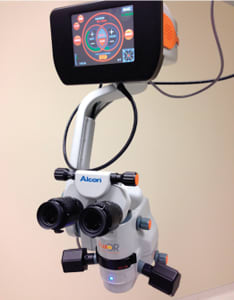
Figure 1: The LuxOR LX3 is part of Alcon’s cataract suite.
Robert Cionni, MD, medical director of the Eye Institute of Utah in Salt Lake City, notes that fewer than 60% of patients who have cataract surgery achieve BCVA within 0.50 D of target while 95% of LASIK patients do. The goal in designing the Cataract Refractive Suite is to achieve cataract outcomes more in line with LASIK, he said. “It’s not just to remove the cataract, but also to get the cataract safely out with a good plan with an excellent refractive outcome for the patient,” he says.
FDA OKs Allegretto excimer
Alcon also announced the FDA approved its Allegretto Wave Eye-Q excimer laser for topography-guided laser treatments used in conjunction with the WaveLight Allegro Topolyzer and topography-guided treatment planning software. WaveLight users will also be able to tailor ablations based on the topography of each patient’s eye.
The multicenter phase III trial on which the FDA based its approval showed that 92.7% of all studied eyes achieved UCVA of 20/20 or better, and 69% achieved UCVA of 20/16 three months after surgery.
“Topography-guided LASIK is an invaluable tool in our refractive armamentarium, allowing us to accurately measure corneal topography and address corneal irregularities as part of the individualized laser treatment,” says chief trial investigator R. Doyle Stulting, MD, PhD, director of the Stulting Research Center and past president of ASCRS.
“Topography-guided LASIK is an invaluable tool in our refractive armamentarium” - R. Doyle Stulting, MD
Nidek adds CATz
Nidek introduced the recently FDA-approved Customized Aspheric Treatment Zone (CATz) for the NAVEX EC-5000 Excimer Laser System (Figure 2), for topography-assisted LASIK procedures. The CATz treatment utilizes the NIDEK OPD-Scan to provide customized correction. The device is designed to be capable of treating myopic astigmatism even in patients with corneal irregularities.
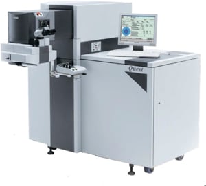
Figure 2: The Nidek NAVEX EC-5000 Excimer Laser with CATz.
PATTERN SCANNING LASERS
Lumenis introduced the Array LaserLink, a pattern-scanning device, days after it received FDA clearance for multi-spot laser photocoagulation. It uses shorter pulse duration and shorter laser sessions than previous platforms to reduce patient discomfort.
The PASCAL Synthesis (Figure 3) retinal laser was on display as part of Topcon’s multi-product launch. The dual-port, pattern-scanning retinal laser is available in 532 nm and 577 nm wavelengths and can be integrated with the Topcon SL-D7 as well as Haag-Streit style slit lamps.
Ellex debuted its Tango SLT and YAG laser and its line of Solo SLT laser systems. The Tango features a dual-mode laser cavity integrated into the slit lamp. It also features a photo-disruptor mode for low-energy, optical breakdown for capsulotomy and iridotomy procedures. The Solo SLT is compact for office space efficiency and features a firing rate of 3 Hz.
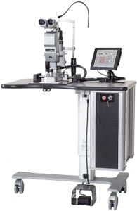
Figure 3: Topcon’s PASCAL Synthesis retinal laser.
DIAGNOSTICS AND IMAGING
OCT and retinal cameras
Optovue debuted its Avanti RTVue XR Widefield Enface OCT, which provides 40° of scanning at 70,000 A-scans per second. The Avanti can provide assessments of peripheral retina pathology, image the optic nerve head and ganglion cell complex in glaucoma, and corneal and anterior segment images and measurements.
Carl Zeiss Meditec demonstrated the Cirrus Photo 600, an integrated system combining a full mydriatic and non-mydriatic true color camera, OCT and optional fundus autofluorescence.
Oculus Surgical presented its Biom Ready single-use wide-angle viewing system, designed for retinal imaging (Figure 4). The Biom Ready includes the Biom HD Disposable Lens.
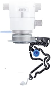
Figure 4: The BIOM Ready single-use wide-angle viewing system.
3-D imaging
Among the multitude of new imaging devices featured at AAO was the Haag-Streit MIOS 5 HD platform, which gives the user the ability to record HD videos using a Haag-Streit surgical microscope.
Haag-Streit also presented a line of surgical microscopes that can integrate Sony Electronic’s medical-grade 3-D video cameras (Figure 5). The technology, specifically designed for ophthalmology, allows 3-D display in the operating room in real-time with a playback option.
Independent of the MIOS 5, the Sony MCC-3000MT 3-D camera system provides both 3-D and 2-D video and can be mounted on most microscopes to capture, record and display 3-D video. It also enables 3-D procedure sharing. Sony also debuted its LMD-3251MT 3-D, 32-inch monitor and ELO LCD 3-D touch screen panel.
The Haag-Streit Octopus 600 (Figure 6), the latest addition to the line, uses flicker stimulus for early glaucoma detection and pathology progression.
Topcon debuts a dozen
Topcon Medical Systems launched nearly a dozen new diagnostic products (Figure 7) at the meeting, among them:
- The 3-D OCT-1 Maestro retinal camera which can scan both eyes and produce an OCT scan and a “true color” fundus image simultaneously. It can also provide red-free images.
- The DRI OCT-1 Atlantis swept-source OCT for posterior imaging, which scans at a speed of 100,000 A-scans/second and allows visualization of the vitreous and choroid in one scan.
- The SP-1P Specular Microscope that can acquire endothelial cell images in seconds with a wide angle “panorama” mode.
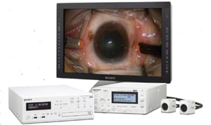
Figure 5: Sony’s medical-grade 3D video system for ophthalmology.
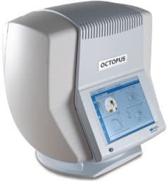
Figure 6: Haag-Streit’s Octopus 600.
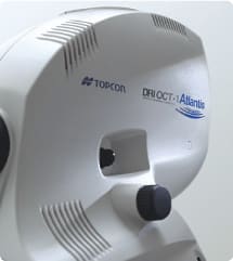
Figure 7: Topcon’s 3D OCT-1 Maestro is a retinal camera/OCT platform.
While these items are not yet available in the United States, Topcon demonstrated several items that are available. Among them, the IMAGEnet 5, a computerized digital system, which can transfer images culled from different Topcon devices.
Topcon’s Synergy Ophthalmic Data Management System provides a secure, remotely accessible, digital archiving for images and other data from more than 130 different devices. It also integrates with most EHR systems.
SURGICAL INSTRUMENTS
Made especially for intraoperative procedures such as microinvasive glaucoma surgery (MIGS), Volk Optical’s TVG (Figure 8) surgical gonio lens is equipped with a floating lens feature, a stabilization ring and multiple pivot points. It can be steam sterilized and can withstand repeated sterilizations.
Accutome presented its Accusharp guarded blades (Figure 9). The angled slit, crescent and straight blade handle designs have the same diameter as unguarded knives, but with thick handles to accommodate the safety guard.
Iridex exhibited its single-use membrane scraper for retinal surgery, available in 23 and 25 gauges. The device is designed to maximize visualization against the retina. Iridex also debuted a portfolio of single-use 27-gauge laser probes compatible with the growing number of 27-gauge vitrectomy systems.
REFRACTION AND MORE
Marco displayed its full Xfraction system, which combines two devices, the OPD-Scan III wavefront technology (Figure 10) and the TRS-5100 automated digital refraction.
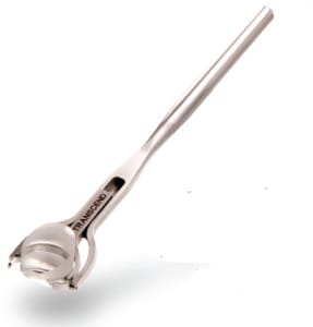
Figure 8: The Volk TVG gonio lens has a floating lens and other features.
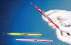
Figure 9: Accutome’s Accusharp guarded blades come in three designs.
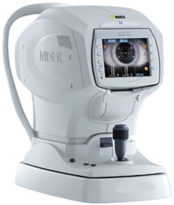
Figure 10: The OPD-Scan III is one half of Marco’s Xfraction system
The OPD-Scan III charts aberrometry data and, within 10 seconds, collects more than 20 diagnostic images. Among its abilities, according to Cynthia Matossian, MD, of Matossian Eye Associates in Doylestown, Pa., is the ability to “get a day Rx and a night Rx with the photopic and mesopic pupil refractions.”
Adds Kerry Solomon, MD of Carolina Eyecare Physicians in South Carolina: “The OPD-Scan III gives you all the information you need under one platform for toric IOLs; accurate keratometry, auto-refraction and corneal topography to determine where the astigmatism lies. At the time of surgery, you also have photographic evidence of pre-op images and postop images to follow overtime. If the lens is going to rotate or move, you will know it with the OPD III.”
The TRS-5100 is a digital refraction system designed to help patients see the difference between lenses more precisely than with traditional methods. It receives data from the OPD-Scan III and completes both WF and AR patient refractions in rapid order. The TRS-5100 eliminates the need for traditional lens dialing tests and questions such as, ‘which is better, one or two?’
Topcon displayed its MC-4S 3-D mirror chart designed to allow for refractions in small areas.
Reichert Technologies featured its all-in-one, customizable digital refraction product, The ClearChart 3P Polarized Digital Acuity System.
Vmax Vision demonstrated its PSF Integra complete refraction lane-in-a-box, which eliminates the need for a 14-to-18 foot refraction lane.
Keeler USA exhibited its 40H slit lamp and its Cryomatic MKII and D-KAT digital tonometer.
WOUND-HEALING AID
Bio-Tissue (Figure 11) debuted its next generation of corneal wound-healing devices, the Prokera SLIM and Prokera PLUS, which reduce inflammation and promote scar-free healing of ocular surface tissue. The Prokera SLIM includes ComfortRING Technology that contours to the ocular surface and moves with the eye to maximize amniotic membrane contact with the cornea and limbus.
The Prokera PLUS incorporates multiple layers of amniotic membrane for durability, making it useful in severe indications such as chemical burns, Stevens Johnson syndrome and corneal ulcers.
TEST KIT AND TOPICALS
At the AAO, Nicox began taking orders for its test kit for the early detection of Sjögren’s, called simply Sjö (Figure 12).
Jason Menzo, Nicox president, says the test utilizes seven biomarkers altogether, including three previously undiscovered ones. Nicox has started distribution of test kits to 600 physicians in 12 markets and plans a full national launch in April 2014. Physicians who want test kits may obtain them by contacting the company, Mr. Menzo adds. Doctors obtain test kits at no cost, and the testing laboratory handles reimbursement and billing.
Bausch + Lomb exhibited Prolensa (Figure 13) for inflammation and ocular pain after cataract surgery, and Lotemax, a topical corticosteroid for postoperative inflammation and pain.
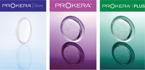
Figure 11: Prokera SLIM and Prokera PLUS were introduced.
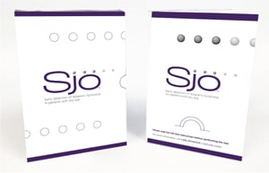
Figure 12: Sjö from Nicox is for early detection of Sjögren’s.
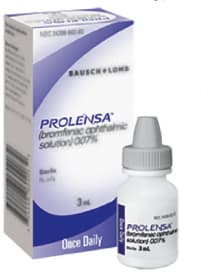
Figure 13: Prolensa, from B + L, is a topical corticosteroid.
EHR AND PACS SOFTWARE
ManagementPlus exhibited its Retina Specific EHR with templates designed to improve efficiency by offering a streamlined, specialty-specific user interface tailored for retinal disease management.
First-Insight presented its cloud-based MaximEyes EHR Software with its interoperability capabilities using the Integrating the Healthcare Enterprise Technical Framework.
NextGen recently updated its Patient Portal to meet Meaningful Use Phase 2 requirements.
Canon debuted imageSPECTRUM version 5.0 image management software to pair with its existing retinal cameras. OM


