Ocular Tissue Donation: Restoring Sight and Advancing Research
The Lions Eye Institute model for tissue research opens up clinical and product development opportunities for academia and industry.
By Lewis R. Groden, MD
Eye banks were first established to facilitate the harvesting and processing of donor corneas for transplantation. The eye banking model has been very successful: corneal transplant is one of the most frequently performed human transplant procedures. Since 1961, more than 700,000 corneal transplants have been performed, with more than 90% of these procedures successfully restoring the recipient's vision, according to the Eye Bank Association of America (www.restoresight.org).
But the role of eye banks — and the need for ocular tissue — now far surpasses simply the processing of whole corneas. With newer transplant procedures, surgeons are now demanding discrete layers of corneal tissue for endothelial rather than full-thickness keratoplasty, specialty femtosecond-laser cut buttons and more. We routinely supply preserved sclera for use in glaucoma valve surgery or repairing ruptured globes with structurally supportive tectonic surgery.
In addition, scientific research that was once conducted mainly on whole corneas or globes is now far more specialized. A glaucoma researcher may need optic nerves from specific demographic groups or a scientist may request just endothelial cells for lab research, for example. A single donor may be able to provide corneas for two corneal transplants, scleral tissue for glaucoma surgery, and retinas, lenses, optic nerves and other tissues for multiple research studies. This new environment is an exciting one that brings both new challenges and new opportunities for eye banks.
Encouraging Donation
We are fortunate that ocular tissue is relatively widely available in the United States, in part because many of the barriers to donation of other organs don't apply to the eyes. Because the cornea is avascular and immunologically privileged, it does not have to be tissue-typed, for example Donation is not disfiguring, which is reassuring to families. Most importantly of all, there are relatively few conditions that prevent donation of ocular tissue.

Figure 1. A donor cornea is evaluated in the slit lamp. COURTESY OF THE LION'S EYE INSTITUTE FOR TRANSPLANT & RESEARCH
There is a serious misperception among the general public — and even among eyecare professionals — that donors must be young and healthy. Studies have shown that donor age isn't a factor in the success of a transplant. Eyes with cataract, glaucoma and macular degeneration are certainly needed for research and are often suitable for transplant. For example, a 70-year-old with AMD may still have a perfectly functioning endothelial pump, making the cornea suitable for transplant, while the retina may be useful for AMD research.
Many conditions — including corneal scarring, HIV/AIDS, hepatitis, prion-related diseases and disorders of unexplained etiology such as Alzheimer's — prevent the use of a cornea for human transplantation; however, there are still many research possibilities for such corneas. Prior history of LASIK may exclude certain kinds of transplants, but previous eye surgery does not, in general, prevent donation for transplantation.
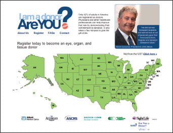
| Are You a Donor? | |
|---|---|
| That's the provocative question posed by SightLife, a nonprofit that seeks to encourage ophthalmologists to sign up to be organ and tissue donors themselves — and, in the process, inspire their patients to do the same. At the recent ASCRS conference, SightLife launched the “Are You a Donor?” campaign, which garnered support from prominent industry players including AMO, Bausch + Lomb, the Allergan Foundation, ASCRS and the Eye Bank Association of America. AMO encourages others in the ophthalmology community to also join in this extremely beneficial program. “We saw an opportunity to partner with SightLife to engage physicians in promoting eye, organ and tissue donation and affirming their personal commitment to help those in need,” commented Jim Mazzo, President of Abbott Medical Optics. To learn more, visit areyouadonor.org. 
| |

Figure 2. Prepping the tissue to begin the pre-cut procedure. COURTESY OF THE LION'S EYE INSTITUTE FOR TRANSPLANT & RESEARCH
Over the years, organ procurement groups and eye banks have learned how to make the donation experience positive and hassle-free for donor families, hospitals and funeral homes.
However, there are steps that ophthalmology can take to encourage more organ donation, and thus provide more tissue for transplantation and research. Visiting your local eye bank and understanding the process of donation and recovery (see sidebar, “The Path of a Donor Eye”) so that you can answer patient questions is an important first step.
A very simple way to raise the level of public awareness about cornea donation is to place local eye bank brochures in your office, and to let patients know that ocular pathologies and procedures do not exclude them from being organ donors. When they are educated about cornea donation, many people — and especially those who have struggled with eye disease or vision problems — truly welcome the opportunity to help others.
Ocular Research
The primary demand for ocular tissue, of course, is still for corneas needed for transplantation procedures. But eyes that are unsuitable for transplant — or portions that remain after a successful corneal transplant — still serve a very important function. Research on human eyes is a critical step in understanding disease etiology and progression, and in the development of new treatments. In most cases, there is no acceptable alternative to human tissue, which means that studies must be delayed or cancelled when human eyes are not available for research.
The Lion's Eye Institute for Transplant & Research (LEITR) is the largest eye bank in the world. We have provided corneas, lenses, sclera, retinas/RPE, trabecular meshwork, optic nerves, endothelial cells and whole globes for a wide variety of research uses. In addition, we can segment donor eyes by disease category, offering an unsurpassed level of low post mortem time and tissue quality for high-demand areas of clinical and scientific research, including macular degeneration, diabetic retinopathy and glaucoma.
LEITR recently opened the world's first ocular research facility to be built in association with an eye bank, at its Tampa headquarters. This is a cutting-edge ocular research center with advanced equipment and 12,000 square feet of research lab space. To support LEITR in this new undertaking, a world-class advisory board has been formed, including the following:
• As chair, Prof. Hank Edelhauser (cornea and drug delivery) from Atlanta's Emory University
• Dr. Jerry Chader (retina) from the Doheny Eye Institute of Los Angeles
• Dr. Abe Clarke (glaucoma) of North Texas Eye Research Institute
• Dr. Mark Petrash (cataract) from the University of Colorado
This unique model allows academic, pharmaceutical and medical device researchers to conduct on-site evaluations and initial preservation of both healthy and diseased tissue for basic science. The ability to conduct research on-site reduces the post-mortem time interval, yielding better experimental results. We anticipate that it will drive the development of new diagnostic tests and pharmacologic treatments for delaying, arresting and preventing progression of ocular diseases.
Examples of retinal basic science that this permits clinical and academic institutions the unique opportunity to pursue include:
• understand the anatomy and components of drusen
• study Bruch's membrane (BM) in dry and wet AMD
• evaluate AMD eyes to modify their BM for study of RPE cell attachment
• RPE cell culture — do they grow like normal RPE cells?
• retinal ganglion cell research
As another example, early-stage or start-up companies at the pre-clinical phase can now use human ocular tissue for product development, as well as testing drugs, drug delivery technology and medical devices, including diagnostics. This could lead to faster and more economical R&D development timelines.
For the future, the possibility of gene sequencing using the LEITR donor database is real, and has already gained some level of interest.
Donations of ocular tissue remain a vital need in ophthalmology. The gift of sight through corneal transplantation has a tremendous impact on patients' productivity, quality of life and personal satisfaction. It's also one of the most rewarding procedures we can perform as ophthalmologists. And every donor cornea or globe offers opportunities for research to expand our profession and our ability to continue offering sight-saving therapies and surgeries to patients worldwide. OM
For more on LEITR, visit www.lionseyeinstitute.org.
| The Path of a Donor Eye |
|---|
| • Hospital or funeral home contacts eye bank upon death of an individual who meets preliminary criteria for donation • Eye bank contacts next of kin (often done in concert with other organ procurement organizations so that the family receives only one call and can grant permission for recovery of many organs/tissues) ► Obtain signed consent ► Obtain medical and social history ► Obtain relevant records from hospital regarding illness and death ► Obtain blood sample • Eye bank creates a donor profile and determines suitability ► Cause of death ► Any medications administered prior to death ► Blood loss at time of death ► Prior disease, surgeries and medications ► Physical signs of infectious disease or intravenous drug use ► Blood sample results (especially for HIV, hepatitis and syphilis) • Recovery of tissue as a whole globe or cornea • Tissue transported to local eye bank • Inspection of tissue ► Microscopic evaluation ■ Cell count ■ Cell morphology ■ Tissue clarity • Final determination of suitability for transplant/research • Placed in cold storage for up to 14 days post-recovery • Surgeon/researcher contacts local eye bank for donor tissue • Tissue provided by local eye bank or elsewhere through nationwide network ► Transplant tissue ■ Matched by age (ideally) to help ensure that the cornea will last throughout the patient's lifetime ■ Prepared according to surgeon request ■ Unused portions of the tissue (sclera, for example) may be stored for other surgical/research use ► Corneas unsuitable for transplant are prepared for research studies, per investigators' request |

|
Lewis R. Groden, MD, is executive medical director of LasikPlus Vision Centers/LCA Vision. He also is an associate professor of ophthalmology at the University of South Florida, codirector of its residency program and medical director at Lions Eye Institute for Transplant & Research. Contact him at lewisgroden@gmail.com. |


