OCT in the Comprehensive Ophthalmology Practice
From preoperative cataract assessment to glaucoma management and detection of retinal disease, SD-OCT has an important role in general practice.

By Amin Ashrafzadeh, MD
Cataract surgery is the bread and butter of a comprehensive ophthalmology practice, and the Cirrus HD-OCT is a valuable diagnostic tool to help us select good candidates for premium IOLs. Being able to recognize an epiretinal membrane (ERM) or vitreomacular traction or other retinal pathologies prior to IOL implantation is critical. Without that due diligence, outcomes can be less than desirable. Patients can have poor vision, and their results may fall short of their expectations. As we know, it takes a thousand happy patients to build a practice, but only one or two angry patients to ruin one.
Preoperative Cataract Evaluation
Not only does the Cirrus HD-OCT give me the data I need, it also helps me educate my patients. If I have a patient with a mild ERM, for example, I can show him the SD-OCT scan, explain what an ERM is and offer my opinion about how it may affect his vision. This allows the patient to make an informed decision whether or not to proceed with premium IOL implantation. This education prior to surgery stages the expectation and can make the difference between a satisfied patient and a dissatisfied patient.
I typically use the macular cube scan for my cataract patients. With it, I can evaluate not only the central fovea but also the surrounding area, enabling me to detect pathologies that are present outside of the central fixation point. For example, a patient's fovea may appear normal, but when I look at an adjacent area on the macular cube scan, I may see clinically significant macular edema from diabetes (see “Macular Edema Uncovered”). There are also times when an ERM may affect the periphery of the fovea more than it does the center.
Looking at the central scan only, it is possible to miss the area adjacent to the fovea, which may be wrinkled and could have a significant pucker that was not previously detected. Cirrus gives me the ability to scroll through the macular cube scan, so I can easily see the area that surrounds the fovea.
Another feature of the Cirrus that I appreciate, particularly when patients have had difficulty fixating, is the Fovea Finder, which automatically identifies the fovea by the reduced reflectivity below the retina. The analysis is then centered precisely on the fovea.
Glaucoma Management
About a third of my practice is comprised of patients who have glaucoma or are glaucoma suspects. With the Cirrus HD-OCT, I can analyze the optic nerve head, the nerve fiber layer, compare the two eyes, and better evaluate the status of the glaucoma.
The Cirrus software is exquisite. It has a progression analysis algorithm, so that I can see how a patient's status has changed over time and how it compares to the normative database.
Evolution of Retina Care
When I started my practice in 2002, I performed all of the focal laser treatments, all of he panretinal photocoagulation, and all of the fluorescein angiograms. The retinal subspecialty has changed dramatically in the past 10 years with the advent of intravitreal injections of various agents. These changes have resulted in significant referral pattern alterations. I now refer patients to retinal specialists earlier because they are better able to serve the patients with newer modalities, such as intravitreal injections. Treatment advances as well as improved diagnostic technology, such as the Cirrus HD-OCT, have changed the relationship between referring optometists, comprehensive ophthalmologists and retina specialists. I believe our referrals and responses are much more accurate and informed. I also believe that referred patients have a better understanding of their conditions and what they can expect from the retina specialist.
Professional Tools
Professional tools are necessary for professional services, whether we are talking about tile work, woodworking or ocular surgery. SD-OCT is a major advancement in retinal and optic nerve head imaging. Cirrus HD-OCT's software and hardware design allow for easy, efficient and accurate acquisition and interpretation of data. ■
| Speed, Simplicity and Improved Patient Flow |
|---|
| We have had the Cirrus HD-OCT in our practice since 2008. Going from the previous generation OCT technology to the Cirrus was like going from a Yugo to a Ferrari. Acquisition speed and accuracy are significantly improved, as is the image quality. The addition of the macular cube scan has taken us from simply looking at a linear foveal image to being able to evaluate a much larger area more precisely and in 3-D. Patient flow has also improved in our office with the addition of the Cirrus. Previously, only one of our technicians was trained with sufficient mastery to regularly operate our OCT machine. It was a chore. The patient had to be positioned properly, and the scan had to be done correctly or it would have to be repeated. In addition, that one technician was the person in our office who took all of our axial measurements. Consequently, patients sometimes had to wait to have their studies done, and when that technician was out of the office, we were in a bind. We used to schedule patients' visits around the availability of the OCT technician, often asking patients to make a separate appointment for their OCT scans and visual fields. That became a prohibitive factor. The Cirrus is so easy to operate that any of our technicians can use it and acquire high-quality scans. It allows quick, easy access, and all the data I need is acquired within minutes. |
| Macular Edema Uncovered |
|---|
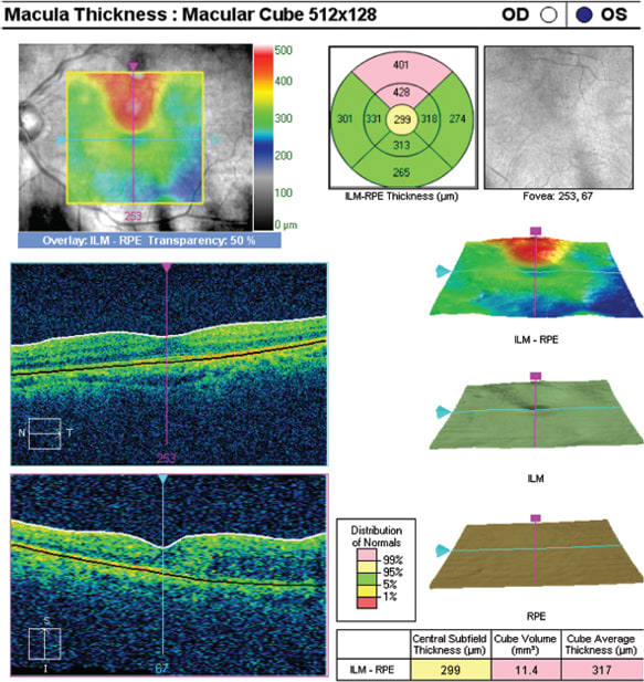
A 51-year-old man with a 10-year history of diabetes and hypertension was evaluated for decreased vision (20/40 OU). Clinical examination showed the patient had moderate diabetic retinopathy with clinically significant macular edema of the left eye. On SD-OCT, the horizontal scan through the fovea is normal. The vertical scan shows some areas of edema. At a very quick glance, the ILM-RPE Overlay image shows the areas of abnormality. The macular cube in this case was helpful in that it allowed a detailed review, even after the patient had left the office, without disruption of the flow of the clinic. The patient was referred to a retinal specialist for further treatment.
|
| When Space Is Finite |
|---|
| One important issue—one that is rarely discussed—is real estate as it relates to equipment. Real estate becomes an expensive commodity very quickly, as most of us are not in a position to expand the size of our practice space. Yet when we make a commitment, often a significant financial commitment, to stay current with our equipment, we must be cognizant of our real space. In our practice of two ophthalmologists, for example, we have 3,400 square feet and in just 10 years, we have acquired a corneal topographer, an anterior segment OCT machine, an IOLMaster, a Cirrus HD-OCT, a second visual field analyzer, and a specular microscope. These are not instruments we can live without, but all of these machines require real estate. The question becomes: With so many instruments and finite space, where do you put them? One of the advantages of the Cirrus HD-OCT is that it is a small box. It requires very little real estate, partly because it is designed for side-by-side viewing. The previous generation OCT required nearly three times as much working space because the operator had to sit behind the instrument rather than on the side of the instrument. Another instrument we tried had a huge footprint, making it impossible to fit into our practice. On the other hand, the Cirrus was a very easy instrument to integrate. Another advantage of side-by-side viewing is that I can help elderly patients who tend to fall back out of the machine. Being able to sit beside the patient while using the Cirrus allows me to use my left hand to help the patient maintain his head position and fixate while I acquire the image. That is a tremendous advantage in that I acquire stable images with very few repeat scans necessary. Additionally, the patient can look at my screen as I interpret the scans for him. The Cirrus is simple, easy to use and compact enough for any practice. |
Amin Ashrafzadeh, MD, is a cataract, cornea and refractive surgeon in private practice in Modesto and Turlock, Calif. He is a consultant for Carl Zeiss Meditec. He can be reached at DrAsh@ModestoEyeCenter.com.


