Anti-VEGF Therapy and Glaucoma
Another productive use for bevacizumab.
MICHAEL B. HORSLEY, MD AND MALIK Y. KAHOOK, MD
Neovascular glaucoma (NVG) is a serious ocular disease associated with elevated intraocular pressure, pain and often poor visual outcomes. Ischemic diseases associated with NVG include proliferative diabetic retinopathy, central retinal vein occlusions, and chronic retinal detachments. In the later stages of neovascular glaucoma, iris and angle neovascularization can lead to the development of peripheral anterior synechiae, which can occlude the drainage system of the eye, resulting in elevated IOP that responds poorly to traditional glaucoma drop therapy and often requires surgical intervention.
This article will discuss usage of bevacizumab in both NVG and as a wound modulator in trabeculectomy.
NVG and anti-VEGF Agents
VEGF is upregulated under conditions of retinal ischemia and neovascular glaucoma. Therefore, it was natural for glaucoma specialists to begin to use anti-VEGF agents for the treatment of NVG. There have been multiple case series that highlight regression of neovascularization of both the iris and angle with the use of VEGF inhibitors.
We first reported a patient with NVG treated with intravitreal bevacizumab after having failed IOP control with transscleral cycloablation as well as panretinal photocoagulation.1 This patient showed an immediate decrease in IOP and improvement in symptoms. Since that time, we now routinely use intravitreal bevacizumab injections to treat patients with NVG and combine therapy with panretinal photocoagulation to decrease the constant neovascular drive that originates from the ischemic retina. We also utilize bevacizumab prior to going to the OR for trabeculectomy or glaucoma drainage-device implantation in patients with NVG to control bleeding, which can be quite extensive in this group of patients.
The use of anti-VEFG agents for NVG has proven quite successful with the following surgical technique.2 After a proper informed consent has been completed, a topical anesthetic, such as tetracaine, is instilled on the affected eye and a lid speculum is put in place. A cotton tip applicator is subsequently soaked with anesthetic and placed over the injection site. Four drops of fluoroquinolone are placed on the eye followed by a drop of povidone iodine. The caliper is set to 3.5 mm for a pseudophakic patient or 4.0 mm for a phakic patient at the inferotemporal limbal border. The syringe is filled with 1.25 mg of bevacizumab (0.05 ml of 25 mg/ml) and the 30-gauge needle is directed toward the mid-vitreous cavity through the site predetermined by the caliper measurements. Then, the plunger is advanced for complete delivery of the bevacizumab aliquot.
A cotton tip applicator is then placed over the site of injection and the needle is removed from the eye to help decrease the possibility of medication reflux. We then place another drop of fluoroquinolone and the speculum is removed. Finally, both IOP and vision are compared with pre-injection levels and an antibiotic is continued four times per day for a total of four days.
Anti-VEGF as Wound Modulators in Trabeculectomy
Trabeculectomy surgery involves the creation of a fistula between the anterior chamber and the sub-Tenon's space to decrease IOP in cases of glaucoma refractory to other therapies. A major barrier to successful trabeculectomy surgery is the body's natural tendency to heal at any incisional site. Fibroblast proliferation with eventual scar formation often leads to occlusion of the fistula and subsequent elevation in IOP. Mitomycin C and 5-Fluorouracil (5-FU), have been used to decrease fibroblast proliferation but often lead to complications such as thin cystic blebs and higher rates of endophthalmitis. The use of anti-VEFG agents as wound modulators for trabeculectomy surgery has been proposed. Since the wound-healing process involves both fibroblast proliferation and angiogenesis, the potential use of these agents seems logical.

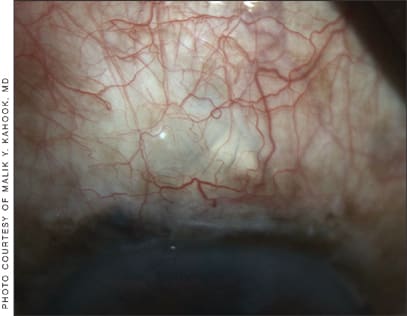
Hypervascular blebs may benefit from treatment with anti-VEGF agents.
We first described the use of bevacizumab during bleb needling to modulate wound healing and noted a significant decrease in IOP that was associated with reduction in conjunctival vessels.3 One milligram of bevacizumab was injected adjacent to the bleb at the end of the needling procedure and resulted in a more diffuse and sustained bleb without complications. Other reports followed that described the utility of bevacizumab and ranibizumab as sub-Tenon's injections at time of filtration surgery or with bleb needle revision. Further prospective studies are underway at the University of Colorado to better understand how anti-VEGF agents might benefit patients undergoing glaucoma trabeculectomy and glaucoma drainage-device surgery. Further studies are also needed to investigate the use of combination therapy (i.e., 5-FU + bevacizumab) for wound modulation post glaucoma surgery since there is no reason to believe that a single agent might result in better outcomes compared to using combination therapy to target multiple pathways associated with wound modulation.
As described previously, the use of needle bleb revisions with bevacizumab is reserved for patients with a previous failed filtering procedure due to excessive fibroblastic proliferation.4 The first goal to ensure a successful bleb revision is an appropriate level of patient comfort.
We instill a judicious amount of lidocaine gel 2% in both the superior and inferior fornices. Then, a drop of fluoroquinolone is instilled. The patient is then positioned at the slit lamp. A lid speculum is used to ensure adequate conjunctival exposure. A Tuberculin syringe with 1.0 to 1.25 mg of bevacizumab (0.04-0.05 ml of 25 mg/ml) with a 27-gauge needle are appropriate for this procedure. The tip of the 27-gauge is bent to a 45-degree angle to improve access to the peripheral portion of the bleb.
Access to the bleb is obtained by placing the needle immediately under the conjunctiva temporal to the area of scarring. Using the needle tip, the fibrovascular tissue is then elevated from the sclera and dissected where possible. Then, the needle is partially withdrawn and redirected to an area temporal to the bleb site in the sub-Tenon's space and bevacizumab is then injected in a depot fashion. A topical antibiotic is again instilled and then continued qid for a total of four days. The patient is reevaluated in one to three days.
Conclusion
The antiangiogenic and antiproliferative effects of anti-VEGF agents have led to studies investigating their utility for treating NVG as well as wound modulation after filtration surgery. Prospective studies are underway to show the utility of these agents and to outline proper treatment methodologies. Research on anti-VEGF agents for various forms of glaucoma may lead to an improvement in IOP lowering without compromising patient safety. OM
References
- Kahook MY, Shuman JS, Noecker RJ. Intravitreal bevaciumab in a patient with neovascular glaucoma. Ophthalmic Surg Lasers Imaging 2006;37:144-146.
- Kahook MY, Olson JL. Treating neovascular glaucoma with intravitreal bevacizumab. Techniques in Ophthalmology 2007;5(4):150-153.
- Kahook MY, Schuman JS, Noecker RJ. Needle bleb revision of encapsulated filtering bleb with bevacizumab. Ophthalmic Surg Lasers Imaging. 2006 Mar-Apr;37(2):148-150.
- Kahook, MY. Needle bleb revision with bevacizumab. Techniques in Ophthalmology 2008;6(4):111-113.

|
Malik Y. Kahook, MD, is associate professor, Glaucoma and Cataract, director, Clinical Research, director, Glaucoma Service, Rocky Mountain Lions Eye Institute, University of Colorado Denver School of Medicine. Michael B. Horsley, MD, is chief resident in ophthalmology at the University of Colorado Denver School of Medicine. |


