Detect Early Glaucoma With Greater Precision
The unique features and capabilities of Cirrus HD-OCT enable you to diagnose glaucoma more accurately.

By Lawrence Stone, MD
Recently, we added Cirrus HD-OCT (Carl Zeiss Meditec, Dublin, Calif.) to our armamentarium to detect early glaucoma.
We've been thrilled with this diagnostic tool. It's been immensely helpful in the decision-making paradigm for patients with ocular hypertension, large cup-to-disk ratios and those whom we suspect may have glaucoma. Over time, I've gained confidence with Cirrus HD-OCT and now find that it's as useful as a 90 diopter optic disk examination of the optic nerve head in assessing glaucomatous damage — although Cirrus HD-OCT focuses more on the retinal nerve fiber layer (RNFL) instead of optic disk cupping.
Cirrus HD-OCT tests for glaucoma using a cube scan, measuring a 6 mm × 6 mm area. It uses a 200 × 200 testing strategy (200 B-scans with 200 A-scans per B-scan) and delivers 27,000 A-scans per second. Because of its fast acquisition time and improved optics, patients with pupils that are 2.5 mm or larger with clear media don't need to be dilated.
The scan is user- and patient-friendly. In 3 minutes, you can prepare the patient, scan both eyes and print the analysis, which is something patients appreciate. The ergonomically designed unit allows the technician to sit on the same side as the patient, enabling him to view the patient's head and body directly.
Scan Readout
The Cirrus HD-OCT glaucoma scan features an RNFL thickness map, an RNFL thickness deviation map, an RNFL TSNIT normative database and an extracted RNFL tomogram graph. The normative database is for the overall RNFL thickness map and the TSNIT.
The RNFL thickness map is the centerpiece of the readout. The RNFL layer is segmented in the 6 mm × 6 mm cube, and the display schematically shows the nerve fiber layer's pattern and thickness (Figure 1). This is the image to show patients, because it's the easiest for them to understand. For comparison, I keep a scan of a normal eye in a folder in each exam room.

Figure 1. The RNFL thickness map schematically shows the nerve fiber layer's pattern and thickness.
The RNFL thickness deviation map displays OCT fundus images (Figure 2). This is an en face view of the cube of OCT data. A colored overlay shows areas of RNFL thickness that deviate from the norm, based on the age-matched normative database. Deviations found in this area are of clinical significance if they're extensions of deviations within the circle. Otherwise, in my experience, they're not indicative of glaucoma.

Figure 2. The RNFL thickness deviation map displays OCT fundus images.
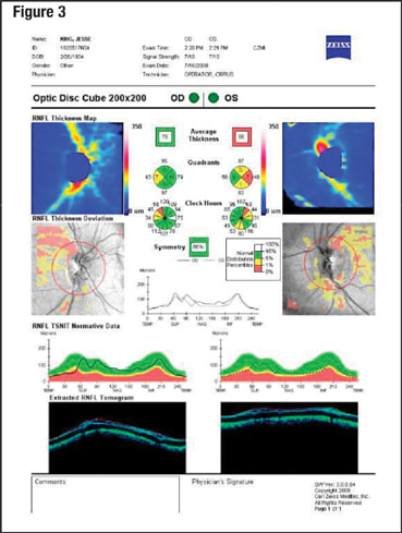
An auto-centration feature identifies the center of the cup and makes a 1.73-mm radius circle around it. The circle is shown as a red line superimposed on the deviation map. The software identifies the center of the disk automatically post-acquisition. Because the center will be in the same place in a repeat scan, no technician-related error (improperly outlining the optic nerve head) can occur. From this 3.4-mm diameter circle, the RNFL tomogram is extracted and forms the basis for the TSNIT analysis. This is compared to the normative database and is displayed graphically (Figure 3).

Figure 3. From the 3.4-mm diameter circle, the RNFL tomogram map is extracted and forms the basis for the TSNIT analysis.
Monitoring for Progression
Detecting significant change in the RNFL over time involves the ability to differentiate between true glaucomatous change and test variability that's not due to glaucomatous damage.1
A recent study2 by Leung and colleagues compared time domain and spectral domain OCT in 35 healthy subjects. The spectral domain OCT demonstrated better measurement repeatability than time domain OCT for total and regional macular thicknesses. This enhanced repeatability indicates that spectral domain OCT, such as Cirrus HD-OCT, would be more effective than time domain OCT in detecting glaucomatous progression. A study3 of Cirrus HD-OCT showed repeatability of scans within a standard deviation of 1.4 microns. The research concluded that the auto-centration feature provides accurate alignment between scans.
As mentioned, Cirrus HD-OCT uses a laser scanning ophthalmoscopic image (LSO) to view the desired fundus image. The LSO image serves as "proxy" for the corresponding "ready to be captured" OCT optic disk data. Once the LSO image is in focus, the technician can click the acquire scan function and capture the HD-OCT scan.
The Cirrus has a repeat mode for monitoring glaucomatous progression, which enables repeat visualization of relevant anatomy. In this mode, you can recall chin and head rest settings of the baseline scan. In addition, you can recall the OCT fundus image of the previous scan (overlay image) and place it in front of the new LSO image (back dropped image).
| A recent study2 by Leung and colleagues compared time domain OCT and spectral domain OCT in 35 heathy subjects. The spectral domain OCT demonstrated better measurement repeatability than time domain OCT. This enhanced repeatability indicates that spectral domain OCT, such as Cirrus HD-OCT, would be more effective than time domain OCT in detecting glaucomatous progression. |
If patient fixation is poor, you can line up the two fundus images in real time and use blood vessels as landmarks as you navigate the point and drag feature. In patients with normal fixation, the autocentration feature provides excellent registration and repeatability.
Prior iterations of LSO or OCT technology didn't allow for registration of anatomical markers from the initial to subsequent studies.
In our private practice setting, we have yet to have the opportunity to follow patients for long intervals. Nonetheless, with the robust technology and future software capabilities of Cirrus HD-OCT, I expect that it will assume an important role in monitoring glaucomatous progression. OM
| References |
|---|
|
| Diagnosing a Glaucoma Suspect |
|---|
A 75-year-old white woman had been coming to see me for several years with IOPs in the high teens. Her GDX scans were normal — although slight asymmetry existed between her eyes — as well as her Humphrey visual fields. Cup-to-disk ratios, documented by stereo disk photography, showed vertical cupping in her right eye at 0.65 and in her left eye at 0.50. The patient's IOP readings were 20 mm Hg in her right eye and 19 mm Hg in her left eye. Her visual acuities with pinhole measured 20/30 OD and 20/30 OS. Mild cortical haze was present in both crystalline lenses. 

Negative Findings The depression in the double frequency visual field was nonspecific and could be attributed to early cataract or other factors. The nerve fiber loss seen on the Cirrus HD-OCT didn't correlate with areas of retinal rim thinning, nor was there glaucomatous visual field loss. Cataractous changes (reflected in the scanning signal strength of the Cirrus HD-OCT) may have accounted for the nerve fiber changes. The comparison of the 90 diopter exams with stereo photos from 2006 reinforced my assessment that the patient didn't have structural glaucomatous changes. This case reminds us that OCT results must be interpreted within the context of other clinical information. A 12-month follow-up visit with this patient, however, is warranted. ■ |
| Low Cup-to-disk Ratio Doesn't Discount Glaucoma |
|---|
A 74-year-old black man presented with decreased vision and was told he might have cataracts. The patient's visual acuities with pinhole were 20/20 in both eyes. His intraocular pressures by applanation were 25 mm Hg OU. The patient's pachymetry readings were 602 microns in the right eye and 597 microns in the left eye. Cup-to-disk ratios were 0.2 in both eyes (Figure 1 and Figure 2). The size of the patient's optic nerve in the right eye was 2.28 mm; the size of the optic nerve in the left eye was 2.18 mm. 

The patient's lenses were clear bilaterally. The Zeiss Matrix 24-2 FDT threshold test showed an inferior hemifield defect (inferonasal step and inferior arcuate) in the right eye and a superior hemifield depression in the left eye. The Cirrus HD-OCT showed a significant superior-temporal nerve fiber defect in the right eye. The left eye showed a generalized depression of the RNFL (Figure 3). The defects were clinically significant when compared with the normative database. 
Final Analysis Although the patient had a low cup-to-disk-ratio, he was diagnosed with glaucoma, which would have been missed by cupping analysis alone. His elevated IOPs, visual field changes and RNFL loss as demonstrated by Cirrus HD-OCT all point to glaucoma. The inferonasal step in the right eye corresponds to superotemporal nerve fiber bundle thinning, and the mild visual changes in the left eye were consistent with the RNFL thickness deviation pattern. To slow glaucoma progression, I prescribed latanoprost ophthalmic solution 0.005% (Xalatan, Pfizer Inc., New York, N.Y.) ■ |


