Perfecting Your Protocol for Infection Prophylaxis
BY MARK PACKER, M.D., F.A.C.S., RICHARD S. HOFFMAN, M.D. AND I. HOWARD FINE, M.D.
Perfection, of course, will forever remain an inspiring yet elusive goal. The random nature of human actions and environmental conditions will, at times, defeat even the most rigorous approach and impeccable technique. Nevertheless, we continue to strive for perfection because of the professional responsibility we have chosen as physicians and surgeons, and because we believe in the benefits of scientific progress.
The state of the art of infection prophylaxis in cataract and refractive surgery continues to evolve, and therefore the standard of care remains a moving target. A plethora of reports has appeared in the scientific literature that surgeons must weigh and consider. As a starting point, it is critical to realize that any data analysis should take into account the multifactorial pathogenesis of postoperative infection. Studies that retrospectively review a case series may fall easy prey to narrative fallacy and confounding errors. As David Chang, M.D. recently pointed out, "… we must be cautious about making practice recommendations based solely on retrospective population studies with multiple covariables."1
Evaluating Risk
In the wake of the European Society of Cataract and Refractive Surgeons' multicenter study of endophthalmitis, the Cataract Clinical Committee of the American Society of Cataract and Refractive Surgery (ASCRS) performed an on-line survey to characterize current practices among members.2 Of the 1,312 respondents, 90% reported an infection rate of less than 1 in 1,000. In general, the published incidence of endophthalmitis after cataract surgery in the peer-reviewed literature ranges from a low of less than 1 in 5,0003 to a high of about 3 in 1,000.4 The risk of severe visual loss from endophthalmitis following cataract surgery has been put at 1 in 6,000.5 For LASIK, the risk of infectious keratitis has been reported to be about 1 in 3,000.6
For cataract surgery, factors that increase the risk of postoperative infection have been identified. Oliver Schein, M.D., M.P.H., has pointed out that "consistent findings have been excess risk associated with corneal incisions, age (especially over 80 years) and loss of posterior capsular integrity…"7 He notes that the "modest" increased risk associated with corneal incisions "can be mitigated by expertise (i.e., close attention to wound construction and integrity)." This statement echoes closely what we have said in the past.8
How to Reduce Risk of Infection
In terms of prophylaxis, several approaches have demonstrated reduced risk of infection. The use of povidone-iodine antisepsis has probably received the most universal support.9 Chemoprophylaxis has demonstrated efficacy by various routes, and is widely employed. In the ASCRS survey, 88% of surgeons used preoperative topical antibiotics and 98% used postoperative topical antibiotics. Intracameral antibiotic administration also received support: 30% of surgeons reported using this route either via irrigation or by direct injection. Subconjunctival administration can also be effective, although it is perhaps less appealing to surgeons performing clear corneal surgery with topical anesthesia due to the stinging and pain it may engender.10 By whatever method, killing bacteria on the ocular surface and inside the eye has value in the prevention of infection.
Assessing Your Approach
In 2004, David Allen, B.Sc., F.R.C. Ophth, and his colleagues presented a series of seven cases of endophthalmitis that occurred in a single surgeon's practice during a 27-week period.11 This surgeon's incidence of infection rose precipitously to 1.6%. At this point, the surgeon stopped operating. Statistical analysis suggested that these cases represented a true outbreak. After examining a variety of potential causative factors, including the timing of cases, nursing staff, equipment, patient risk factors and microbiology, investigators determined that the "only common contributory factor in each case was the surgeon."
A review of the surgeon's technique determined that 2 weeks prior to the occurrence of the first case, he had discontinued his prior practice of administering a subconjunctival antibiotic injection at the conclusion of surgery. Following this analysis, the surgeon resumed his use of subconjunctival antibiotic injections; he enjoyed a zero incidence of endophthalmitis in the subsequent 1,350 cataract operations he performed.
| Sample Protocol Outline | |
|---|---|
|

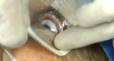
Figure 1. Following sterile preparation of the skin the upper eyelid is retracted with a 1″x 5″ suture strip. The strip is placed as close to the eyelid margin as possible. 
Figure 2. The upper and lower eyelashes are covered with one Tegaderm, cut in half. 
Figure 3. The operative eye is covered with an ophthalmic drape. This drape is fenestrated and has an attached fluid collection pouch. |
This cautionary tale demonstrates how a systematic investigation into an outbreak of infection led to the correct causative factor. When infection strikes, surgeons should first determine if the event represents a random and statistically expected event; if it does not, investigation is warranted to determine the cause. Casting a wide net by reviewing all the potentially relevant factors in an outbreak of infection increases the likelihood of finding the culprit. This approach is analogous to that undertaken in looking for the source of outbreaks of non-infectious postoperative inflammation, such as toxic anterior segment syndrome (TASS)12 and diffuse lamellar keratitis (DLK).13
Significance of Results
Assigning the correct significance to the results of published studies as they apply to local conditions represents a second important lesson from this paper. At the time, subconjunctival injections were deemed "possibly relevant but not definitely related to clinical outcome" according to an evidence-based update by Thomas Ciulla, M.D.9 However, an earlier report had suggested a possible link.14 Nevertheless, the key to understanding the outbreak lies in the fact the subconjunctival injection represented the only antibiotic prophylaxis this surgeon employed. Discontinuing the injection meant dispensing with all chemoprophylaxis. In a different setting, for example, one where the surgeon uses topical antibiotics both pre- and postoperatively, the discontinuation of subconjunctival antibiotics might not be significant.
Recently, Ng et al. performed a retrospective study of endophthalmitis in Western Australia from 1980 to 2000 by examining 205 cases of postoperative infection and four time-matched randomly selected controls for each case. The authors found a significant impact from antiseptic preparation and subconjunctival antibiotic injection. Interestingly, antisepsis was nearly universal in both cases and controls while subconjunctival injection was about 50/50 in controls and 30/70 in cases. Postoperative topical antibiotics were also nearly universal in both groups, while intracameral antibiotics were rare in both groups. Ironically, the power of the study to detect a significant difference for subconjunctival injections was higher than its power to detect a difference for antisepsis or topical antibiotics. The fact that it did still find a difference for antisepsis confirms again the importance of povidone-iodine; the fact that it did not find a difference for topical antibiotics does not mean they are worthless.
Turning to wound location, Ng et al. did not find a significant difference between cases and controls based on scleral, limbal or clear corneal incisions. In a recent thorough review of the literature, Mats Lundstrom, M.D., concluded that, "There is no conclusive evidence of the relationship between clear corneal incision and endophthalmitis."15 Nevertheless, clear corneal incision design and construction appear to be less forgiving than scleral tunnel incisions.16 When reviewing potential causes for infection, a leaky wound with hypotony represents a clear avenue for introduction of bacteria into the eye. The use of correct architecture and the attainment of a Seidel negative closure are critical for the prevention of infection (Figure 4).17 A single suture should be placed if necessary. Incision construction represents an important area for examination when faced with an increased incidence of infection or if self-sealing cannot be routinely obtained.

Figure 4.OCT of the anterior segment (Visante, Carl Zeiss Meditec, Dublin, Calif.) demonstrates the profile of a temporal clear corneal incision constructed with the 3D Trapezoidal Diamond (Rhein Medical, Tampa, Fl.). The incision is constructed by placing the tip of the blade just anterior to the corneal vascular arcade and directing the knife towards the corneal apex in a single planar motion. The differentially beveled blade is designed to create a tunnel that is 2 mm in chord length.
Preventing Infection, Step by Step
Infectious disease subspecialists generally consider pathogenesis in terms of both the resistance of the host and the virulence of the etiologic agent. Most cases of post-surgical endophthalmitis are related to normal flora of the eyelids and ocular surface, for example, staphylococcus epidermidis. Gram-negative organisms, such as pseudomonas aeruginosa, though capable of more rapid destruction of tissue, occur less frequently. While the etiology remains fairly consistent, host factors may vary widely. The most significant factor remains the patient's age. However, thorough examination of the ocular adnexa, with special attention to signs of blepharitis, eyelid malposition and lacrimal insufficiency or obstruction, forms an important step in assessing infection risk. Initial treatment with eyelid hygiene for blepharitis or definitive surgical treatment of ectropion or nasolacrimal duct obstruction makes sense prior to cataract or refractive lens surgery. Less obvious, treatment of dry eye syndrome (e.g., with topical cyclosporine) prior to surgery may improve the quality of the tear film and strengthen its defensive mechanisms. The presence of poor or partial blinking due to a facial palsy may indicate a need for increased lubrication with artificial tears in the perioperative period. Consideration of host factors such as these may lead to special precautions in the setting of reduced resistance to infection.

Figure 5. Seidel test is performed at the conclusion of surgery after the intraocular pressure has been adjusted to a physiologic level. If persistent leakage occurs, stromal hydration is performed. Massage of the corneal surface with the side of a cannula sometimes facilitates apposition of the roof and floor of the incision, leading to sealing. Occasionally a 10-0 nylon suture is required to effect closure.
Preoperative antibiotic prophylaxis to sterilize the ocular surface remains a mainstay of infection prophylaxis. Many surgeons begin topical antibiotics, especially a fourth-generation fluoroquinolone, moxifloxacin or gatifloxacin, up to 3 days prior to surgery. Additional topical antibiotic is often administered in the immediate preoperative period along with mydriatic agents and non-steroidal anti-inflammatory drops. Topical antibiotic administration is frequently continued following surgery for a period of 1 to 2 weeks. Some surgeons have adopted perioperative oral antibiotic prophylaxis, as fluoroquinolones exhibit excellent penetration into the vitreous body.
Sterile preparation of the eye for surgery forms the most critical aspect of infection prophylaxis, as has been shown by the nearly universal adoption of povidone-iodine. Important considerations for draping include optimal ergonomic access to the eye, complete sequestration of the lids and lashes and maintenance of fluid drainage away from the surgical field. Critical aspects of intraoperative technique include incision construction and avoidance of complications. In particular, compromise of the capsular bag is recognized as posing increased risk of infection. Surgeons should consider the use of intracameral antibiotic agents, either in the irrigation solution or as an injection into the anterior chamber. At the conclusion of surgery the incision, whether clear corneal, limbal or scleral, must be checked for watertight closure following adjustment of the intraocular pressure to physiologic levels (Figure 5). A leaky incision requires stromal hydration, massage or suture closure. Removal of the lid speculum and drapes should be accomplished without putting pressure on the eye. The postoperative exam is performed 2 to 24 hours after surgery, and the patient is instructed to call the doctor immediately if there is increased pain and decreased vision.
A Careful, Critical Eye
A surgeon's primary challenge in preventing endophthalmitis consists of keeping a critical perspective on infection prophylaxis. We hear many reports and see multiple studies, some of which contain relevant information and provide thoughtful insights. We should always evaluate the conclusions and opinions of others in light of our own experience. The best way to build knowledge is to document outcomes; as human beings we are generally too much concerned with the results of our most recent or most unusual experiences. Documenting outcomes and tracking one's own data allows an objective measure of risk as well as the potential to improve results by following up on alterations in protocol and technique after they are made. OM
References
- Chang DF. Reducing the risk of endophthalmitis after cataract surgery. J Cataract Refract Surg. 2007;12:2008-2009; author reply 2009.
- Chang DF, Braga-Mele R, Mamalis N, Masket S, Miller KM, Nichamin LD, Packard RB, Packer M; ASCRS Cataract Clinical Committee. Prophylaxis of postoperative endophthalmitis after cataract surgery: results of the 2007 ASCRS member survey. J Cataract Refract Surg. 2007; 10:1801-1805.
- Buzard K, Liapis S. Prevention of endophthalmitis. J Cataract Refract Surg. 2004;9:1953-1959.
- Nichamin LD, Chang DF, Johnson SH, Mamalis N, Masket S, Packard RB, Rosenthal KJ; American Society of Cataract and Refractive Surgery Cataract Clinical Committee. ASCRS White Paper: What is the association between clear corneal cataract incisions and postoperative endophthalmitis? JJ Cataract Refract Surg. 2006;9:1556-1559.
- Lundström M, Wejde G, Stenevi U, Thorburn W, Montan P. Endophthalmitis after cataract surgery: a nationwide prospective study evaluating incidence in relation to incision type and location. Ophthalmology. 2007;5:866-870.
- Solomon R, Donnenfeld ED, Azar DT, Holland EJ, Palmon FR, Pflugfelder SC, Rubenstein JB. Infectious keratitis after laser in situ keratomileusis: results of an ASCRS survey. J Cataract Refract Surg. 2003;10:2001-2006.
- Schein OD. Prevention of endophthalmitis after cataract surgery: making the most of the evidence. Ophthalmology. 2007;5:831-832.
- Fine IH. "Clear corneal cataract incisions require attention to detail," in Packer M, Hoffman RS, "Cataract Corner." Ophthalmology Times. 2003;2: 12-13
- Ciulla TA, Starr MB, Masket S. Bacterial endophthalmitis prophylaxis for cataract surgery: an evidence-based update. Ophthalmology. 2002;1:13-24.
- Ng JQ, Morlet N, Bulsara MK, Semmens JB. Reducing the risk for endophthalmitis after cataract surgery: population-based nested case-control study: endophthalmitis population study of Western Australia sixth report. J Cataract Refract Surg. 2007;2:269-280.
- Mandal K, Hildreth A, Farrow M, Allen D. Investigation into postoperative endophthalmitis and lessons learned. J Cataract Refract Surg. 2004;9:1960-1965.
- Mamalis N, Edelhauser HF, Dawson DG, Chew J, LeBoyer RM, Werner L. Toxic anterior segment syndrome. J Cataract Refract Surg. 2006:2:324-333.
- Hoffman RS, Fine IH, Packer M, Reynolds TP, Bebber CV. Surgical glove-associated diffuse lamellar keratitis. Cornea. 2005;6:699-704.
- Lehmann OJ, Roberts CJ, Ikram K, Campbell MJ, McGill JI. Association between nonadministration of subconjunctival cefuroxime and postoperative endophthalmitis. J Cataract Refract Surg. 1997;6:889-893.
- Lundström M. Endophthalmitis and incision construction. Curr Opin Ophthalmol. 2006;1:68-71.
- Masket S. Is there a relationship between clear corneal cataract incisions and endophthalmitis? J Cataract Refract Surg. 2005;31:643–645
- Fine IH, Hoffman RS, Packer M. Profile of clear corneal cataract incisions demonstrated by ocular coherence tomography. J Cataract Refract Surg. 2007;1:94-97.
| Mark Packer, M.D., F.A.C.S. is a clinical associate professor at the Casey Eye Institute at Oregon Health & Science University in Portland, I. Howard Fine, M.D., is a clinical professor, and Richard S. Hoffman, M.D., is a clinical associate professor. The doctors are principals in the practice, Drs. Fine, Hoffman & Packer, LLC, located in Eugene, Ore. |

|


