Going Digital Makes a Great Impact in Daily Practice
The EyeRoute system allows you to analyze patient data fast, which would be difficult or impossible to do using film or paper.

By John Parkinson, M.D.
Implementing an integrated digital image network can revolutionize the way you practice and provide patient care. At Four Corners Eye Clinic in Durango, Colo., we've been using the EyeRoute ophthalmic image management system (Anka Systems Inc./Topcon Medical Systems Inc.) for the past several years. Before implementing the system, we'd been considering how we might convert to electronic medical records (EMR). We realized fairly quickly that converting to EMR would present challenges. The charting functions of the EMR packages we looked at weren't very user-friendly. Also, it was clear we'd have a difficult time getting the four doctors in our practice to agree on a product.
However, when we became aware of the EyeRoute ophthalmic image management system, we had no problem agreeing to purchase it. While we would've been able to get images from our diagnostic equipment into an EMR system, our ability to manipulate, display and analyze data and images would've been limited.
With the EyeRoute, we're able to integrate images and reports from various diagnostic instruments into a single, secure digital environment. This is important because I specialize in glaucoma and do a fair amount of retina care. The ability to efficiently access and view patients' past and present visual fields, OCT scans and other test results has an enormous, positive impact in daily practice.
Furthermore, EyeRoute was a great step toward EMR. We have extensive medical records on many patients, and the longer we use EyeRoute, the easier the conversion will be to full EMR. Here, I explain how I use EyeRoute to facilitate efficiency and provide quality patient care every day.
Instant Access
In our office, we have a 20-inch, high-resolution monitor on each exam room counter. Before I walk into the room, a technician accesses the patient's file so that the tests performed that day are ready for viewing. When a file isn't open, the screen is either locked for HIPAA compliance purposes, or we run one of several educational videos for the patient to see. As soon as I walk into the room, I click on the patient's test results so we can review the findings together. If at any time I also want to view the patient's past test results, I simply click on what I want to see and it appears.
Our EyeRoute system is structured so that we can scan images into the system. For example, we can scan old 35-mm optic disc photographs, so they're instantly available to me, along with all of the data imported directly from our instruments. We also can scan Polaroid photographs or other information we receive from other doctors' offices. And we can scan printed results from our corneal topographer right into the system. Finally, we can add external photos, such as those related to ptosis and blepharoplasty, from the memory card of a digital camera.
Viewing and Analyzing Patient Data
Having all of a patient's test results at my finger-tips via EyeRoute is a real time-saver. Just as important, it allows me to view patient data in a variety of ways. For instance, I can access a basic screen that lists all of the patients I've seen in one day. Or, I can search for a particular patient by typing any three letters of the first or last name. Clicking on the patient's file displays a list of all images in the EyeRoute system for that patient. At a glance, I can see which tests have been done, when they were done and for what tests the patient is due.

Figure 1. EyeRoute allows the physician to compare data over time in several different ways. This view shows a patient's fundus photographs taken several years apart.

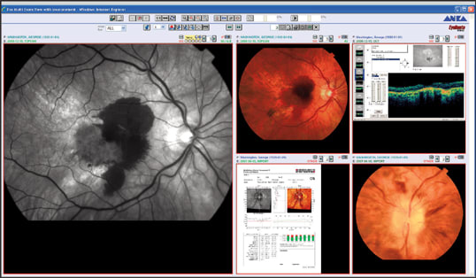
Figure 2. The multi-exams function (shown here) is a useful way to compare test results over time.
The following are some of the many ways I use EyeRoute to view and analyze patient data.
■ View multiple images at once. In the upper right hand corner of the screen, I can click on an icon to simultaneously view any two or any four of a patient's images.
■ Compare test results from different visits. There are several different ways to look at data over time, which is one of the strengths of the EyeRoute system. For example, with a few clicks of the mouse, I can view a patient's fundus photographs from an exam in 2005 on the left side of the screen and fundus photographs from an exam earlier this year on the right (Figure 1).
■ Magnify images. Clicking on the magnification tool enables me to zoom in on any portion of an image. I often use this feature with my glaucoma patients to look for changes in the contour of blood vessels on the optic nerve or to closely examine signs of retinal pathology.
■ Contrast enhancement. This feature is especially helpful when I want to take another look at one of the older photographs that I've scanned into EyeRoute. Even if the image quality isn't ideal, I can use the contrast tool to get a clear view.
■ Compare test results over time. Clicking the "multi-exams" button is another useful way to compare test results over time. I often look at visual fields this way. I click multi exams to access all of a patient's visual field results for both eyes. I can hit the "single pan" button, which allows me to scroll through any of the tests to compare the right eye over time and the left eye over time. I can look at the grey scale and the statistical plots as well. I also can use multi exams to compare different tests over time (Figure 2), such as fundus photographs, OCT scans and angiograms.
■ Compare baseline to subsequent tests. The new Humphrey GPA (glaucoma progression analysis) software (Carl Zeiss Meditec) merges nicely with EyeRoute's capabilities. The analysis software enables me to view all of a patient's previous visual fields on one screen. With this information imported into EyeRoute, I can click to show the right or left eye, keeping the baseline test on the left, which shows the mean deficit of all fields that have been performed in the past. Then, by simply clicking the right side, I can view today's visual fields and all previous fields up to that point. It's a great tool for showing patients whether or not there's been glaucomatous progression.
■ Correlate two different testing modalities. Another view I use can place fluorescein angiograms on the same screen with OCT scans, so I can determine how the findings correlate.

Figure 3. In the exam room, the physician can use the EyeRoute to easily view a selected sampling of a patient's fluorescein angiography and OCT scans over time.
■ Sampling over time. Many of our patients with age-related macular degeneration have had multiple injections of anti-vascular endothelial growth factor agents. In these cases, it's helpful to use EyeRoute to access their last four tests, such as OCT scans and fluorescein angiograms, and view them on the same screen. I can show patients how they've responded to treatment with regard to the amount of macular edema, subretinal or sub-RPE fluid present. It also may be useful to know how the current test compares with previous tests. The simple way to do that is to skip a few tests by clicking the box for each of the tests I want to see, creating a kind of random sampling over time (Figure 3).
■ Historical view. To get a complete historical view, simply click the multi exams button. This displays all of the tests that we've performed for the patient. But remember that on most exam dates, more than one scan from each test was sent into EyeRoute. So if I designate a certain number of columns per line, I can see that number of scans, from every exam through time, across the screen. This shows me in an instant how the patient's condition has progressed over time.
■ Designate key images. I use this function when I see patients by clicking to "designate and save" only the most important images. Then, when I search all of the data over time, only the key images come up. By doing this, I build a repository of key images and eliminate those that aren't important for future patient care. It's a simple and effective way to clean out the database.
As you might imagine, it would be very difficult to do this kind of work with a paper or film system, but the EyeRoute makes it easy.
Other Useful Capabilities
I've also found the EyeRoute system to be a great team-building tool for our referral relationships. I can save all of the images from EyeRoute as jpeg files and e-mail them, or cut and paste pictures to be used in letters that I send to referral sources. Even more helpful, when I want a referring doctor to be involved in a patient's care, I simply use the EyeRoute interface to assign the doctor's name to the case as a referring provider. Then, I can assign the doctor a password and user ID, so he can access our server — whether he has the EyeRoute system or not — and review the patient's test results. Referring doctors enjoy the ability to share test results with their patients in their practices as well. Likewise, if I need to refer a patient to another doctor, rather than packaging printouts of OCT scans, angiograms and other test results, I can give the doctor a password, and he can access the patient's data quickly and easily from his own practice. The response to this has been great.
Ready for the Future
Our practice is completely delighted with the EyeRoute system. Every doctor, from the youngest to the oldest, is using it.
In addition, once we're ready to make the move to full EMR, what we have in place already will be a major benefit. By adding only an electronic charting and note-taking component, we'll have an efficient, functional EMR system that will probably cost less than some of the EMRs we'd priced previously. OM
Dr. Parkinson practices at Four Corners Eye Clinic, a regional eyecare center in Durango, Colo. You can reach him at John@fourcornerseye.com.


