ON TECHNOLOGY & TECHNIQUE
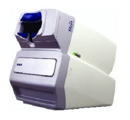
Marco�s RTA 5 Imaging System
The RTA 5 (Retinal Thickness
Analyzer) is a versatile early-detection imaging system offering
complete diagnosis of diabetic retinopathy, AMD, glaucoma and other
retinal pathologies. The RTA 5 is able to assess the disc, macula and
peripapillary regions and is a useful tool in all diagnostic phases �
early detection, diagnosis and follow-up progression analysis from any
field session.

Validity
Clear and straightforward data, provided by two diagnostic pathways �
scanning laser ophthalmoscope and high-resolution digital fundus imaging
� add to the RTA�s value in clinical settings.
The quick and easy non-mydriatic exam (3+ mm pupil) provides:
- high-density thickness maps automatically overlaid on a digital fundus
image
- disc topography maps
- retinal nerve fiber layer (RNFL) cross-section charts
- interactive 3-D imaging and
- deviation probability maps.
Precise registration methodology assures the mapping accuracy necessary
for diagnostic validity.
Fundus Imaging
The RTA�s integrated charge-coupled device camera captures a fundus
image with every scan measurement. The image is captured simultaneously
with retinal and/or disc scans, maximizing patient comfort and practice
efficiency. A 2-D or 3-D retinal thickness map superimposed on the
patient�s fundus image provides benefits important to diabetic
retinopathy screening, as well as other retinal pathologies.
Digital images are captured in 0.33 to 0.48 seconds and a
high-resolution image is displayed immediately on a large screen,
providing an educational tool that can be used during patient
consultations. All digital images are automatically saved in TIF format,
facilitating electronic archival and EMR communications. The RTA�s
automatic registration software makes it possible to combine up to eight
individual images, creating a wide 60�x 72� fundus image.
Dynamic 3D Anatomy Imager
The Dynamic 3D Anatomy Imager creates a full volume, 3-D, cross-section
of a specified area of the retina or disc, automatically overlaid on the
patient�s fundus image. This interactive program enables free movement
and assessment across all grid lines of the scanned retina, without the
need to reposition and retest adjacent locations.
The 3-D images obtained are easy to interpret and measure. Video
captures provide patient education and professional presentation
material. They can also be sent to colleagues and the physician�s
referral base.
Vision Wellness Examination
RTA�s Vision Wellness Examination plays a role in the vision loss
prevention initiative by effectively detecting, identifying and
monitoring the major back-of-the-eye diseases
in their earliest stages. The test is
non-mydriatic and lasts only 2 to
3 minutes. The exam increases patient comfort and compliance by
providing enhanced scheduling flexibility and smooth patient flow.
Applications in Glaucoma
The RTA 5 system provides multiple diagnostic assessments for early
glaucoma detection and management. Most important is the ability to
assess the complete health of the optic nerve head and macula � in every
standard RTA test. Fundus images and real slit-sections are always
integrated with thickness, topography, RNFL and dynamic 3-D-anatomy
imaging modalities.
The full posterior pole is examined with overlapping scanning laser
ophthalmoscope scans, for the most broad and complete screening for
glaucoma. Additionally, user-defined test patterns allow thorough
imaging across the optic nerve head and full peripapillary region.
Traditional rim/cup data and temporal-superior-nasal-inferior-temporal
mapping is provided in each patient session. OM
For more information on the
RTA 5, please contact Marco at (800) 874-5274 or visit
www.marco.com.


