glaucoma
Structural
Analysis:
An Important Aspect of
Glaucoma Assessment
BY LESLIE GOLDBERG, ASSISTANT
EDITOR
|
Case Study |
|
| A practical application of HRT. The following case study is presented by Anne L. Coleman, M.D. The patient is a female, 68 years of age, followed since 1994. | |
|
Initial
Examination: ► Untreated IOP was 23 mm Hg OD and 26 mm
Hg OS |
OS
► The visual field in 1997 had an abnormal
Glaucoma Hemifield Test (GHT) due to several abnormal points on the nasal horizontal
midline |
|
Follow-up Examinations: OD ► The visual field remained consistently
normal until 2006 when a defect in the superior hemifield was detected |
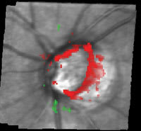
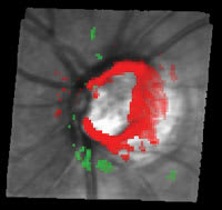
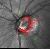
Clinical Impression The HRT detected glaucomatous damage in 1994 while the visual field was still normal. Over the following 12 years, the visual field showed fluctuation but it was not until 2006 that a repeatable visual defect could be confirmed. Progressive structural damage was also detected by the HRT. In this case, the HRT was able to detect damage earlier than conventional methods and also detected progressive structural change before the visual field showed a confirmed defect. |
The clinical emphasis on structure and function evaluation in glaucoma has undergone a transformation in recent years. In the past, glaucoma detection and management relied heavily on functional assessment, typically white-on-white automated visual field tests. However, studies have revealed the potential limitations of relying too heavily on visual field results.1-4
In summary, visual fields can be insensitive for detecting early glaucomatous damage, and they often have a high degree of variability, which limits the clinical usefulness of a single test result. This finding prompted The Ocular Hypertensive Treatment Study (OHTS) researchers to change the study protocol and require three consecutive abnormal visual fields before the patient was said to have converted to glaucoma, which may be impractical for everyday practice.
Emphasis on Structural Assessment
Recent longitudinal studies investigating structural change over time have helped validate the role of structural imaging for glaucoma. Until now, the evidence for structural assessment was based only on cross-sectional studies. In cases where the structure was abnormal but the function was normal, it was unclear whether the structural results were incorrect (false positive), or if the structure was picking up an abnormality missed by the functional test. Only a longitudinal study that follows patients over a long period of time can answer this question.
The OHTS is a multi-center, randomized clinical trial that followed ocular hypertensives longitudinally. The study is well known for its main findings suggesting the therapeutic benefit of lowering IOP in ocular hypertensives as well as its identification of important risk factors for glaucoma. However, the OHTS study also had important implications for understanding the role of structural assessment in glaucoma (See "Implications of OHTS," page 118).
|
|
||

|

|

|
|
|
|
|
|
Figure. The Progression Analysis OS. HRT shows large areas of significant change starting in 2001. |
||
The most powerful predictor of glaucoma was the Moorfields Regression Analysis (MRA), which measures rim area and adjusts for optic disc size to determine if the eye is in the normal range. Remarkably, in some cases, the MRA was abnormal on Heidelberg Retinal Tomography (Heidelberg Engineering, Vista, Calif.) several years before any defect was detected by visual field testing or by evaluation of good quality stereo-photographs by an expert reading center. It is likely that the ocular hypertensives with an abnormal HRT at baseline already in fact had glaucoma, but at enrollment were thought to be normal, based on a normal visual field and a "normal" appearing optic disc.
The OHTS ancillary study found that the HRT could identify patients both at high risk for developing glaucoma and also patients at low risk, with the latter group identified with a greater than 90% accuracy. Anne L. Coleman, M.D., professor of ophthalmology and epidemiology at the Jules Stein Eye Institute at UCLA says, "This is the first study of its kind to suggest that an automated retinal imaging device such as the HRT can predict which ocular hypertensive patients will not develop glaucomatous damage over the next 5 years."
Clinical Applications
Many clinicians are beginning to recognize the potential value of imaging results, such as those obtained with the HRT. It can be difficult, however, when the clinician tries to apply this information from clinical studies to everyday clinical practice. Despite the growing body of evidence suggesting structural assessment is useful in clinical evaluation of a patient, especially from imaging devices such as the HRT, it is clear that this opinion should not be overstated. "Functional tests will always be an integral part of clinical practice. The challenge lies in incorporating information from both structural and functional assessment to form conclusions for the individual patients seen in everyday practice," adds Dr. Coleman.
|
Implications of OHTS |
|
► The study found that
the majority of ocular hypertensives who converted to glaucoma did so based on an
abnormality detected by optic disc assessment, rather than by visual field assessment.
► The OHTS Ancillary included structural imaging with the HRT as part of its protocol. ► The OHTS Ancillary found that numerous HRT measures could predict the development of glaucoma — in some cases several years prior to detectable damage to either the visual field or with clinical optic disc assessment. |
References
1. Quigley HA, Dunkelberger GR, Green WR. Retinal ganglion cell atrophy correlated with automated perimetry in human eyes with glaucoma. Am J Ophthalmol. 1989;107:453-464.
2. Dandona L, Hendrickson A, Quigley HA. Selective effects of experimental glaucoma on axonal transport by retinal ganglion cells to the dorsal lateral geniculate nucleus. Invest Ophthalmol Vis Sci. 1991;32:1593-1599.
3. The Ocular Hypertensive Treatment Study (OHTS) sponsored by the National Institutes of Health.
4. Keltner JL, Johnson CA, Levine RA, Fan J, Cello KE, Kass MA, Gordon MO. Normal Visual Field Test Results Following Glaucomatous Visual Field End Points in the Ocular Hypertension Treatment Study. Arch Ophthalmol. 2005;123:1201-1206.
Go to www.ophmanagement.com/article.aspx?article=86654 to view visual fields for the patient's left eye and visual fields and HRT results for the right eye.


