feature
Surgical Pearls for
the Ultimate
Soft-Shell Technique
Trypan blue is a component of an evolving
surgical method.
BY
STEVE ARSHINOFF, M.D., F.R.C.S.C.

Trypan Blue and the USST
In November of 1999, I became the first Canadian ophthalmologist to use Vision Blue as a component of a variant of my ultimate soft-shell technique (USST). I employ the USST with trypan blue when performing phacoemulsification and IOL implantation in patients with black and white cataracts. Healon5 (Advanced Medical Optics, Inc. [AMO], Santa Ana, Calif.) and balanced salt solution are used as part of the USST to facilitate the cataract procedure and enhance its safety.1 The cohesive viscoadaptive agent fills the anterior chamber, thereby pressurizing the eye and protecting the corneal endothelial cells. Balanced salt solution is used below the viscoadaptive agent, away from the incision, and creates a surgical operating space with low viscosity. This, in turn, makes capsulorrhexis, hydrodissection and later, Healon5 removal, much easier.
|
|
|
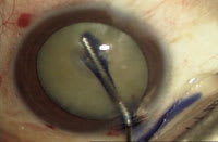
Figure 1. Vision Blue is painted over the anterior capsule. |
Initially, I employed a two-step technique where Vision Blue was substituted for balanced salt solution. I later modified the technique and followed the injection of Vision Blue with pressurization of the anterior chamber using balanced salt solution. Subsequently, I read the publication of the Marques three-step technique that involves the injection of Healon5, followed by balanced salt solution, and then Vision Blue — a reversal of the last two steps of my USST technique. With the USST technique, the initial slow injection of balanced salt solution washes out any excess Vision Blue. This is followed by one rapid, smooth injection of balanced salt solution, which forces the viscoadaptive ophthalmic viscosurgical device ("OVD") mass to move around and block the incision, thereby pressurizing the anterior chamber. Both steps are done as one smooth motion. I find that my technique yields a clearer view of the capsule for performance of capsulorrhexis.2,3
The details of the technique are as follows:
►Fill the anterior chamber 80% to 90% with a viscoadaptive OVD. Be careful not to inject initially at the wound to avoid blocking the incision.
►Next, paint trypan blue over the capsule (Figure 1). Use a tiny drop and eject it onto the capsule surface through a 27-gauge, hockey-stick cannula that is attached to a tuberculin syringe containing trypan blue. Paint the dye over the capsule surface using the distal ''blade'' of the hockey stick.
►Slowly inject balanced salt solution under the viscoadaptive OVD and onto the capsule surface using a 10-cc syringe and a similar 27-gauge, hockey-stick cannula. A slow injection will wash out the excess trypan blue but ensure that the overlying OVD layer will not move.
►Last, increase the injection speed to ''pulse'' and inject balanced salt solution away from the wound, with the cannula aperture positioned on the capsule surface near the pupil margin distal from the incision, in the USST style. This will force the OVD upward and backward to block the incision and pressurize the eye. Figure 2 shows a capsulorrhexis performed with this technique. The view of the capsule is crystal clear, which greatly facilitates capsulorrhexis.
|
|
|
Figure 2. The USST modified for trypan blue. Notice the clear view of the capsule. |
Better, Safer Visualization
The use of this modified USST-trypan blue technique offers a number of advantages. First, minimal trypan blue is needed to obtain optimal capsular staining and other intraocular tissues are not stained. This is significant because trypan blue is listed in the Merck Index as a carcinogen. It is imperative that the intraocular contents are exposed to as little of any foreign substance as possible.
Second, the method provides a completely clear view of the capsule. The presence of a viscoadaptive OVD that blockades the wound allows for excellent pressurization of the anterior chamber and a capsulorrhexis that is easy to perform and is not likely to tear out to the periphery.
Finally, trypan blue decreases capsular elasticity, therefore making pediatric cataract surgery much easier. The sum of all these advantages is that this technique significantly improves the ease and timeframe of the surgical procedure for white, black and pediatric cataracts, and results in fewer complications.
Steve Arshinoff, M.D., F.R.C.S.C., is professor of ophthalmology at the University of Toronto and a surgeon at the Humber River Regional Hospital in Toronto, Ontario, Canada.
References
1. Arshinoff SA. Using BSS with viscoadaptives in the ultimate soft-shell technique. J Cataract Refract Surg. 2002;28:1509-1514.
2. Marques DMV, Marques FF, Osher RH. Three-step technique for staining the anterior lens capsule with indocyanine green or trypan blue. J Cataract Refract Surg. 2004;30:13-16.
3. Arshinoff SA. Letter. Capsular dyes and the USST. J Cataract Refract Surg. 2005;31:259-260.


