spotlight on technologu &
technique
An
Early Predictor of Glaucoma
The
Heidelberg Retina Tomograph 3 measures variability and change over time.
By
Leslie Goldberg, Assistant Editor
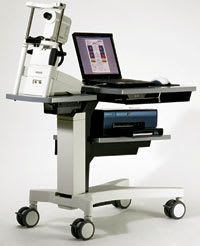
The versatility of Heidelberg's (Vista, Calif.) upgraded HRT, the Heidelberg Retina Tomograph 3 (HRT3), provides physicians with multiple new options and capabilities in the areas of glaucoma, retina and cornea. The HRT3 helps doctors to assess, diagnose and manage glaucoma patients by using laser technology to produce a topographical image of a patient's optic nerve, providing an objective analysis of the structure's cup, rim and retinal nerve fiber layer. The retina software option provides 3-D images of retinal pathology and edema maps that can help to locate and quantify macular edema. The Rostock Cornea Module adds confocal scanning microscopy to the HRT3, allowing for the acquisition of high-resolution images of corneal cell structures with 1-μm resolution.
"The HRT3 is 100% compatible with both the HRT1 and HRT2 data," says Michael Sinai Ph.D., director of Clinical Applications at Heidelberg Engineering. "This is especially valuable to our existing client base of glaucoma doctors who may need to follow a patient for an extended period of time."
|
|
| Heidelberg Engineering's HRT3. |
How the HRT3 Works
The HRT3 scans a patient's eye with a beam of light, producing a topographical or surface image of the optic nerve disc, and then measures the cup, rim and retinal nerve fiber layer. The instrument stores each patient's images and on a follow-up visit, the device checks for signs of statistical change that are then presented to the clinician as possible signs of disease progression. "Any tool that helps us detect progression enables the clinician to make a better decision on a course of therapy," says Robert Fechtner, M.D., professor at the Institute of Ophthalmology and Visual Science, UMDNJ-New Jersey Medical School.
"The HRT3 measures both variability and change over time, allowing progression analysis to be based upon statistical methods. You must know the variability in order to determine if observed change is real disease progression or just random fluctuation. We faced similar problems with visual field analysis in the past. The HRT3 is the first imaging technology to offer this strategy," says Dr. Fechtner.
An Important Finding for Glaucoma Patients
An ancillary study to the NEI-sponsored Ocular Hypertension Treatment Study (OHTS) evaluated the associations between baseline optic-disc measurements obtained using confocal scanning laser ophthalmoscopy (CSLO; Heidelberg Retinal Tomograph) and the development of primary open-angle glaucoma (POAG). The study found that many baseline HRT measurements, when used alone or combined with central cornea thickness, IOP, and history of vascular disease, could predict the development of POAG among individuals with ocular hypertension.1 "This OHTS ancillary study show that the HRT can predict which ocular hypertensive eyes will develop glaucoma prior to visual field defects or detectable damage using stereo-photography of the optic disc," says Dr. Fechtner.
"This is the first longitudinal study of its kind. It suggests that the HRT can help predict or detect damage, sometimes years before conventional methods might," he adds.
Features
The following are new features incorporated into the HRT3:
►The Glaucoma Probability Score. This feature uses an advanced form of artificial intelligence called a relevance vector machine. The analysis provides a statistical probability of glaucoma using ethnic-specific databases. The software analyzes the shape of the patient's anatomical structure using a 3-D model of the optic disc and the peripapillary RNFL. The program then calculates a probability of structural abnormality based on how closely the patient's model compares to healthy and glaucomatous shapes.
►Advanced Glaucoma Analysis. Built upon the previous HRT platform, this tool utilizes a variety of parameters including the Moorefield's Regression Analysis and larger, ethnic-selectable databases to improve diagnostic accuracy.
►Complete Cup, Rim and RNFL Analysis. These analyses provide a comprehensive assessment of the structure. The calculated parameters are adjusted for age, optic disc size, and are compared to the appropriate ethnic database. Any abnormal values are automatically flagged for the physician.
►Optic Disc Size Measurement and Classification. This feature alerts the clinician to unusual disc sizes, which are typically more difficult to diagnose.
►Asymmetry Analysis. By utilizing OU databases to look for OD/OS differences, the clinician can detect suspicious cases when comparing the patient's own eyes to each other instead of relying exclusively on normative data.
►Patient's RNFL Values. The data are plotted while the machine traverses the optic disc, beginning temporally, moving superiorly, nasally and back to temporal (TSNIT). These RNFL values are superimposed on the RNFL normative data in order to detect areas that fall below the normal range.
►Topographic Change Analysis (TCA). This feature provides a statistically based progression algorithm that accurately detects structural change over time. The HRT compares variability between exams and provides a statistical indicator of change. TCA automatically aligns subsequent images with the baseline exam, providing a point-by-point analysis of the optic disc and peripapillary RNFL.
►Active Quality Control Measures. This real-time feedback helps the operator obtain high-quality images. Separate tests for focusing, illumination and optic disc centering give the operator guidance to obtain the best possible images with every scan. Quality is indicated in plain language on every printout ranging from "very good" to "very poor."
►Laptop Technology. The HRT3 is built on laptop technology enabling a small lightweight package for transporting to multiple office locations. A wireless printer provides space-saving flexibility and convenience.
Reference
1. Zangwill LM, Weinreb RN, Beiser JA, et al. Baseline topographic optic disc measurements are associated with the development of primary open-angle glaucoma: the Confocal Scanning Laser Ophthalmoscopy Ancillary Study to the Ocular Hypertension Treatment Study. Arch Ophthalmol. 2005;123(9):1188-1197.


