feature
Cleaner LASIK: Is it Possible? (Part 2)
Eliminating
"exposure-to-closure" complications.
BY
L.C. LAHAYE, M.D., H.H. RIEKE, PH.D. AND F.F. FARSHAD, PH.D.
How much can the innovative design of handheld instruments contribute to minimizing LASIK complications? In light of the authors' experience, the answer is "quite a bit."
Granted there is variability in techniques, outcomes, results and complication rates from surgeon to surgeon. However, innovative handheld instrument design that enhances a surgeon's skill and experience by enabling standardization of technique is the bottom line for achieving reproducible and consistently successful results. In this article, we will emphasize handheld device design.
Comparing Complication Rates
Stage I of the LASIK procedure is simply the keratotomy or "the incision." Improvements in design and manufacture have been responsible for making this aspect of the LASIK procedure straightforward and much safer. Today, the incidence of complications directly associated with the keratotomy is in the range of only 0.1% to 0.3%.1
In Part One, Cleaner LASIK: Is it Possible?, (October 2005) we discussed the potential complications associated with Stage II of the procedure, which begins the moment the corneal flap is reflected and ends with the corneal flap returned and sealed in its original position.
Inspection of the data supports the assertion that the majority of less-than-desired outcomes, complications and other potential problems that continue to take a toll on patients, surgeons and the industry originate from the multitasked Stage II. These problems and their rate of occurrence include:
►infectious lamellar keratitis - 0.3%
►diffuse lamellar keratitis (DLK) - 1% to 4%
►undercorrection and overcorrection - 4.9% to 15%
►decentered and off-axis ablations - < 1.0%
►flap striae - 1%
►epithelial undergrowth/ingrowth - 1.3% to 2.2%
►foreign matter in the flap/bed interface - 3.2%
►nuisance problems and procedural delays (bleeding at the keratotomy site onto the stromal bed/interface, involuntary eye movements, momentary laser interruption and associated problems)
►loss of best-corrected visual acuity from irregular ablations, islands
►toxic plume dispersal and optic splatter
The aggregate data show Stage II complications to be a minimum of 29 times greater than Stage I complications.
|
|
|
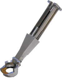
The LaFaci handpiece is ergonomically designed. It downsizes to contain the surgical field. |
A Closer Look at Stage II
There are no less than nine functions (in addition to laser dose delivery) required of Stage II. These include: (1) containment of the surgical field; (2) fixation and control of eye movements; (3) corneal flap placement; (4) removal of beam-masking, flap-bed surface fluid/moisture; (5) plume evacuation; (6) irrigation; (7) aspiration; (8) flap repositioning and realignment and (9) flap adherence. Each step is predisposed to complications. These functions must be executed with exactness, standardization and strict adherence to basic surgical principles and techniques in order to avoid less-than-desired outcomes.
How can we elevate the surgeon's practice and skill level to reduce complications? This appears to be a difficult challenge due to the variability of surgical techniques, accompanying devices and the varying levels of surgeon experience.
LASIK-specific surgical devices, which address one or more of the nine functions, were borrowed from general ophthalmology or developed during the 1990s. These devices were usually designed to perform a single function and include the Thornton fixation ring, aspirating LASIK speculum, microsurgical sponges, assorted flap-irrigation cannula, spatula instruments, flap forceps and sponge ring.
These devices traditionally are limited, require a great deal of manipulation and are somewhat abrasive. They do not downsize or provide for comprehensive containment from incision exposure to closure. They provide marginal flap management and, at best, offer only partial protection against irrigation backwash.
Both the laser and keratome procedures are well defined and standardized. But post-keratotomy flap management, stromal-bed hydration control, minimization of tissue manipulation, maximization of surgical field containment and minimization of irrigation backwash, along with plume evacuation, still remain areas that are suspect in outcomes that require additional surgical procedures, present vision-threatening complications and contribute to a less-than-desirable operating room environment.
An Easier Way
Looking for an easier, more enjoyable and "cleaner" way to perform each step of Stage II LASIK, the first author (Dr. LaHaye) and associates developed the LaFaci Surgical System with Vision Pro, LLC, Lafayette, La.
The LaFaci (LASIK Facilitator) System, an automated device, carries out nine-plus specialized functions through a single, ergonomically styled handpiece (Figure 1), facilitating the entirety of Stage II of the LASIK procedure, thereby allowing for improved technique standardization and reduction of some of the problems discussed. It is somewhat like a Swiss Army knife for LASIK surgery.
The handpiece is placed on the eye prior to lifting the flap and remains positioned until flap fixation. This is fundamental for correct "surgical wound" management.
The functions executed by the LaFaci Surgical System, from surgical incision exposure to closure include: limbal downsizing and surgical field containment; ciliary vessel tamponade; at-the-site plume evacuation; flap management; target-stroma aeration hydration control; on-demand sterile irrigation without concern for backwash; instantaneous aspiration of surgical debris; positive fixation and control of eye movement; flap repositioning and accelerated flap adhesion. These steps are crucial in providing a cleaner, consistent LASIK outcome.
Results and Initial Experience
After receiving Class 2 510 clearance from the FDA for the LaFaci Surgical System in 2004, Dr. LaHaye has used the LaFaci System on a total of 225 eyes. Stage I was accomplished using a standard microkeratome.
Inclusion criteria included eyes correctable to �20/20, a myopic spherical equivalent of less than -11.50 D, and corneal thickness sufficient to maintain at least 270 microns of residual post-ablation bed thickness.
A limitation that exists in the study is the low follow-up. Investigation shows that the patients who did not return were those who were doing well and did not feel that follow up was necessary.
At 3-months and 6-months postop, results on 107 eyes (47.6%) and 85 eyes (37.85) yielded a spherical refractive equivalent mean of -0.16 D, ± 0.30 D, and -0.15 D ± 0.26 D respectively. Eighty-two percent and 84% of the eyes were within ± 0.25 D of intended correction at 3 and 6 months respectively, while 97% and 98% were in ± 0.50 D. All eyes were within ± 1.00 D.
The results showed that UCVA of 20/25 or better was achieved in 93% of patients seen at 6-months postop. Rate of loss of 1 or more lines of BSVA was 5% at 6 months while 12% gained 1 or more lines. Most importantly, no eyes experienced infectious keratitis, DLK, epithelial ingrowth/undergrowth, flap striae, decentered ablations, entrapment of surgical debris in the flap interface, or have required a retreatment for under- or overcorrection.
Epithelial defects did occur in four eyes (1.7%) during Stage I, which did not require aborting Stage II. It was noted flap replacement using LaFaci allowed minimal manipulation and did not extend or enlarge the keratome-created epithelial defects.
Achieving Clean LASIK
There is a learning curve for the LaFaci Surgical System similar to that of phaco. The level of standardization the instrument provides obviates a variety of complications and potential health concerns for patient and surgeon alike.
LASIK is a corneal procedure. It makes little surgical sense to have the ocular adnexa as part of the surgical field, especially since the tissues of LASIK are uniquely avascular and lack "aqueous absolution." By using a handpiece that isolates the surgical field, including the flap, through Stage II, a more sterile procedure is possible.
In addition to limbal downsizing and containment, cleaner LASIK also requires a means of providing sufficient I/A capabilities to thoroughly cleanse and rehydrate without concern for pooling or backwash.
The use of titrated amounts of aspiration and irrigation in sterile manner allows reduced flap manipulation and the potential for visually significant striae. Reducing the number of instruments used, especially the elimination of surgical sponges, can potentially decrease the incidence of epithelial ingrowth and DLK. Enhanced accuracy and efficacy of ablation can be realized from at-the-site removal of beam masking plume, consistent hydration management of the target stroma and surgeon-controlled fixation whether an eyetracker is used or not.2,3
The Goal: Reducing Retreatments
Retreatments are the issue. Retreatments include any procedures required for the correction of flap striae, epithelial ingrowth, under- or overcorrection, decentered ablations, off-axis ablations, DLK, infectious keratitis and other complications.
Retreatments can be costly in terms of lost time and revenue associated with additional surgery, pre- and post-care expenses, and time that could have been spent seeing new, paying patients. Also consider that negative word-of-mouth leads to a decline in refractive volume. Conservatively, the projected financial burden of single retreatment is $3,000. An "only 5%" retreatment rate on 500 eyes costs a practice a minimum of $75,000. Retreatments also take an emotional toll on staff from patient comments like; "Why didn't you get it right the first time?"
The industry has afforded us lasers, keratomes and promises of providing our patients with "super vision". However, these technologies can only go so far in reducing retreatments. It is up to us, as refractive surgeons, to continue to work at providing our patients with consistent improvements. Just as emmetropia was our goal for cataract surgery back in the late 1980s, zero retreatment is now our goal for refractive surgery. In striving to better outcomes and advance safety for our patients, we will uphold and grow their trust and respect.
Leon C. LaHaye, II, M.D., is the medical director of LaHaye Total Eye Care in Lafayette, La. His e-mail is ifxiis@lahayesight.com. Herman H. Rieke, Ph.D., is professor of petroleum engineering at the University of Louisiana at Lafayette. Fred F. Farshad, Ph.D., is a Chevron-endowed research professor in the department of chemical engineering at the University of Louisiana at Lafayette.
References
1. Majmudar, P.A., 2004. LASIK Complications. Focal Points AAO, XXII (13): 1-9
2. Bueeler M. MSE, Mrochen, Limitations of pupil tracking in refractive surgery: systematic error in determination of corneal locations. Refractive Surg, 2004; 20:371-378.
3. Guttman, Lipshitz,. Predictable super vision outcomes may be difficult goal. Ophthalmology Times. 2004; 5:48-50.


