feature
The ExPress Mini Shunt
An Option for Use in
Difficult-to-Control Glaucoma
BY MARLENE R. MOSTER, M.D. AND
ANNA K. JUNK, M.D.
The ExPress mini-glaucoma shunt (Optonol Ltd., Neve Ilan, Israel) is a 400-μm wide by 3-mm long, stainless steel device that offers an additional option when treating difficult glaucoma cases. Currently, when medical and laser therapy has failed, some type of filtration surgery is the next step taken. However, in patients who have already failed one trabeculectomy, or have had multiple other procedures such as corneal grafts, vitrectomies or retinal detachment repair with prior cataract surgery, the options are either to repeat a trabeculectomy or to consider a large tube shunt such as the Baerveldt (AMO, Santa Ana, Calif.), Ahmed (New World Medical Inc., Rancho Cocamonga, Calif.) or Molteno (Molteno Ophthalmic Ltd., Dunedin, New Zealand).
|
|
|
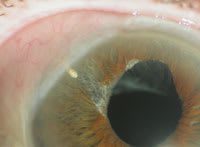
Figure 1. The right eye showing the failed trabeculectomy and superior scar at the 12 o'clock position. |
The ExPress shunt has the advantage of being somewhere in between these two options. It provides immediate consistent aqueous flow through a 50-μm opening that allows for formation of a posterior, low-diffuse bleb usually limited to one quadrant. The bleb formation starts immediately and microcysts within the bleb can often be seen within the first or second post-operative day. This is not always the case with repeat trabeculectomy, since the scleral flap often has to be closed tightly, given the increased chance of hypotony with scarred tissue. Often, the bleb is slow to develop even with mitomycin. With repeat trabeculectomy, there is the concern of cutting sutures too early, which may cause a shallow or flat anterior chamber.
Additionally, the surgery with the ExPress is less time consuming than with a larger shunt, and should it fail, a more extensive shunt procedure can be planned. The down side is that at least a few clock hours of movable, clear conjunctiva must be available for this shunt to work. Unlike the larger tube shunt, this is "conjunctival dependent" in the superior area of the limbus.
Technique with the ExPress
The current technique while using the ExPress
is to place it under a scleral flap. The original technique of placing the shunt
directly under the conjunctiva has largely been abandoned. Placing the shunt under
the flap affords better flow characteristics, less hypotony and also protects both
the shunt itself and the conjunctiva from erosion.
We generally employ two releasable
sutures on either side of a 4x4-mm scleral flap. This provides us with excellent
control of the IOP in the immediate postoperative period. We have found that when
the releasable sutures are placed 1 mm from the limbus through the scleral flap,
the direction of aqueous flow is posterior, which is exactly where we want it.
This allows for formation of a diffuse bleb. The releasable sutures can be removed either individually or together at the slit lamp, depending on the IOP. We generally like to aim for an IOP of around 13-17 mm Hg on the first day, so that the patient's vision will return to baseline quickly.
|
|
|
Figure 2. Eight weeks after the ExPress R-50 shunt. All the sutures from the conjunctiva had been removed at 3 weeks. |
All the sutures, conjunctival and releasable, are usually removed by the third postop week. Patients are placed on prednisolone every 2 hours while awake for the first week and then q.i.d. for the next 5 or 6 weeks. A fluoroquinolone is taken q.i.d. for 1 week, and an NSAID b.i.d. or t.i.d. for the first few weeks, if the cornea does not have superficial punctuate keratitis. If the eye is dry, we avoid NSAID use.
Case Study
An 80-year-old retired health educator who presented in Feb. 22, 2005 to Wills Eye Hospital with an IOP of 36 mm Hg in his right eye while on brimonidine b.i.d, dorzolamide b.i.d and travoprost q.h.s. in both eyes. His vision was 20/25 OD, 20/20 OS. He was healthy except for controlled hypertension.
Past Ocular History
The patient had had pseudoexfoliation since 1992 and was diagnosed, treated and operated on out-of-state. He had argon laser trabeculoplasty (ALT) in 1992 and again in 1998. Both eyes were complicated by a history of iritis. In 2000, he received phaco in his left eye with a posterior IOL, trabeculectomy with mitomycin C (MMC) and an anterior vitrectomy, followed by bleb needling. In 2005, he experienced a sudden loss of vision in his left eye, dislocated lens and a re-sutured IOL to the sclera.
In 2002, he received phaco in his right
eye with posterior IOL and an anterior vitrectomy. He had an IOP of
32 mm Hg
preop and was treated with medications only. In 2003, his right eye underwent a
trabeculectomy with MMC superonasally. He presented with a choroidal detachment
OD, which was subsequently drained. In 2005, the patient's IOP was 40 mm Hg OD and
he was placed on maximum medical therapy for a failed trabeculectomy. The patient
was referred for treatment when his visual field was thought to be progressive.
|
|
|
Figure 3. Sixteen months following superotemporal surgery with the ExPress R-50 shunt at the 10 o'clock position. The bleb is diffuse and posterior. IOP is 15 mm Hg and patient is not on any glaucoma medications. |
Ocular Exam
His visual acuity was 20/25 OD and 20/20 OS. His IOP was 36 mm Hg OD and 19 mm Hg OS. The patient had an afferent pupillary defect in his right eye and pseudoexfoliation involving the anterior segment in both eyes. His anterior chamber had an irregular pupil with some iris atrophy in the right eye. The IOL was partially in the sulcus and partially in the bag. He had a scarred trabeculectomy nasally and superior conjunctival fibrosis at 12 o'clock (Figure 1), with one quadrant of movable conjunctiva superotemporally.
He had a sewn-in posterior chamber lens in his left eye and a minimally functioning trabeculectomy. There was no iritis in either eye. The angle was open 360� with few posterior synechiae from prior laser procedures in both eyes. His optic nerves measured 0.9 mm in the right eye and 0.6 mm in the left eye. His retina was flat with mild cellophane macula changes in the right eye and macula drusen in both eyes.
The patient's visual field had advanced arcuate loss and a nasal step with a small central island remaining in the right eye and mild arcuate loss in the left eye. Heidelberg Retinal Tomograph (Heidelberg Engineering, Inc., Vista Calif.) scans displayed advanced loss of the neural retinal rim 360� in the right eye and mild to moderate changes consistent with glaucoma in the left eye.
Clinical Course
The patient underwent an ExPress R-50 shunt to the right eye on Feb. 24, 2005 with 2 minutes of MMC and two releasable sutures. This was accomplished with a combination of intracameral 1% non-preserved lidocaine, 2% lidocaine jelly and subconjunctival 1% non-preserved lidocaine on a 27-gauge cannula in the superotemporal quadrant, where the conjunctiva was mobile. On the first day postop, IOP was 17 mm Hg and vision 20/70 OD. One week postop, vision was 20/30, IOP was 15 mm Hg and steroids were reduced to q.i.d. Antibiotics were stopped and NSAID decreased to b.i.d.
Eight weeks postop, the patient's vision was 20/25, IOP was 14 mm Hg and all conjunctival and releasable sutures were removed (Figure 2). On June 15, 2006 (16 months post-op), vision was 20/30 and IOP was 15 mm Hg. No glaucoma medicines were used and there was a low, diffuse bleb. The patient had a stable but advanced optic nerve and visual field.(Figure 3)
Conclusion
Patients who present with prior surgical procedures, especially failed trabeculectomies, are difficult to treat. The ExPress R-50 offers the glaucoma surgeon an alternative to either repeating a trabeculectomy or placing a more extensive silicone tube shunt in those patients in whom the IOP has been shown to be higher than the optic nerve can tolerate.
Marlene Moster, MD, is a professor of clinical ophthalmology at Thomas Jefferson Medicine College in Philadelphia, PA. She is an attending surgeon at Wills Eye Hospital. Anna Junk, M.D., is assistant professor at the Bascom Palmer Eye Institute in Miami, Fla.


