feature
Assessing the Value of ECP
Used in conjunction with phaco, ECP may improve the quality of life for patients with cataracts and glaucoma.
BY
JOHN PARKINSON, ASSOCIATE EDITOR
In recent years, a growing number of surgeons have been combining phaco with endoscopic cyclophotocoagulation (ECP). According to those who use the technology, adding ECP to phaco not only lowers IOP, but can also improve patients' quality of life by reducing their number of glaucoma medications.
Surgeons are also replacing phaco/filtering procedures with phaco/ECP in some cases of patients with medically uncontrolled glaucoma and refractory glaucoma. This has led these surgeons to offer the combined procedure to an increasingly larger patient base.
In addition, a major study conducted by Stanley J. Berke, M.D., and colleagues confirmed the combination procedure's efficacy in lowering IOP and reducing the number of medications. (For more about the study, read "The Latest Phaco/ECP Data" on page 20.)
This article will explore the history behind phaco/ECP, the advantages and limitations of the procedure and how anterior segment surgeons are utilizing it in their practices.
|
|
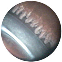
| Dr. Mackool says it is better to have 3 to 4 ciliary processes in camera view as opposed to 1 or 2 so as not to overtreat. |
The Melding of Two Procedures
The development of ECP began after its inventor, Martin Uram, M.D., a retina specialist, became frustrated with the lack of treatments for neovascular glaucoma. At that time, Dr. Uram says the main treatment was cyclocryotherapy, which was painful to the patient, had limited to moderate success in bringing down IOP, and a high complication rate.
Dr. Uram says ophthalmologists knew if they could treat the ciliary processes they would be able to reduce pressures, but issues such as access and visualization remained. "If you could have an endoscope to visualize the ocular interior, you could find your way to the ciliary body and then laser it. You would diminish aqueous fluid production, and the pressure would go down," explains Dr. Uram.
During the development process, Dr. Uram tackled instrumentation size and illumination issues before refining the endoscope to its present form. He also decided to change the location of the incision from the pars plana to the limbus. Dr. Uram later went on to cofound a company (Endo Optiks, Little Silver, N.J.), which now produces the ECP technology.
Performing phaco in conjunction with ECP was first suggested by Richard J. Mackool, M.D., director, Mackool Eye Institute, Astoria, N.Y., who decided to use the combined procedure for his patients with cataracts and medically controlled glaucoma. Dr. Mackool found that by adding ECP, patients enjoyed the benefit of long-term IOP reduction and required fewer medications to maintain these levels over time. Dr. Mackool has been performing phaco/ECP for more than 10 years and estimates he and his associates have done over 1,000 of the combined procedures.
Benefits
|
|
|
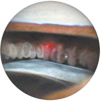
The red spot is the aiming beam of the ECP laser centered squarely on the ciliary process. |
Dr. Mackool says there are a number of reasons ECP works well in conjunction with phaco: both must be performed as intraocular surgery, are utilized with small incisions, use approaches that permits ready access to ciliary processes and are easy and safe to perform by the experienced surgeon.
The phaco/ECP procedure offers many advantages including a reduction in IOP, cataract removal and reduced dependence on glaucoma medications. Dr. Uram considers the reduction of glaucoma medications after surgery the main advantage of phaco/ECP.
"The purpose of the treatment is not to lower the intraocular pressure per se, but to trade off the lower pressure for less meds," acknowledges Dr. Uram.
Dr. Berke says ECP may offer patients more sustained IOP stability than a medication-only regimen. "Some studies have shown that not only the level of the pressure [is important], but that patients who have pressure fluctuations are more prone to have disease progression," he says.
Robert J. Noecker, M.D., vice chair of the Department of Ophthalmology and director of the Glaucoma Service at the University of Pittsburgh, says surgeons can achieve a greater reduction of IOP by performing a more extensive ECP treatment.
"You have more control at the time of the procedure to affect the outcome, so the more you treat [the ciliary processes], the lower the pressure is going to be."
Another benefit of ECP is that a surgeon can utilize the endoscope to monitor the anatomy associated with the phaco aspect of the procedure, such as viewing the condition of the zonules and capsular bags or locating the IOLs' haptics.
Surgeons are also reporting helpful practice management efficiencies with phaco/ECP. This combined technique adds approximately 5 minutes to a traditional phaco, whereas, a phaco/trabeculectomy may add 25-30 minutes of intraoperative time. There is also less follow-up required with phaco/ECP than with phaco/trabeculectomy.
"After a trabeculectomy, a lot of the surgeon's skill is required to keep the bleb functioning," says Dr. Uram.
|
The Latest Phaco/ECP Data |
|
Dr.
Berke and colleagues at Ophthalmic Consultants of Long Island performed a 5-year
study looking at phaco-only procedures compared to phaco/ECP procedures. They performed
the study on patients with medically controlled glaucoma to test the effect on lowering
IOP and reducing the dependence of glaucoma medications. Dr. Berke reported the
results at the American Glaucoma Society earlier this year. In a 707-eye study, Dr. Berke and colleagues performed phaco/ECP on 626 eyes and compared the results to 81 eyes receiving phaco only. They showed the patients receiving the combined procedure achieved a greater IOP reduction postoperatively: 19.08 mm Hg preop [±4.14], 15.73 mm Hg postop [±3], (P<.0001) than did patients with phaco only: 18.6 mm Hg preop [±3.38], 18.93 mm Hg postop [±4.12], (P=.60). Dr. Uram says most phaco-only patients experienced a benefit of a decrease of IOP in the first year postoperatively, but their IOPs gradually elevated. "After the first year, IOP drifted back up so that by the third year it was a little higher than when they started," says Dr. Uram. In comparison, the phaco/ECP group's IOPs decreased and stayed down. A reduction in the number of medications was realized postoperatively in the phaco/ECP patients. This group required fewer medications to maintain lower IOP levels: 1.53 medications preop [±0.89], 0.65 medications postop [±0.95], (P<.0001). There was no reduction in required medications to control IOP in the phaco-only group. These patients used1.20 medications preop [±0.83], 1.20 medications postop [±0.87], (P=0.5). Over the long-term, 68% of patients who underwent combined phaco/ECP required at least one less medication to control IOP postoperatively, while only 11% of the patients who had phaco alone enjoyed this benefit. Lastly, a cost/benefit analysis was done to see what patients could be saving be decreasing one medication each. An average savings of $1,500 a year per patient was calculated for the phaco/ECP group while the phaco-only patients slightly increased their expenditures. In a separate multicenter, retrospective pilot study of resource use and cost associated with glaucoma, physicians reported the cost of treatment ranged from $623 per patients per year for glaucoma suspects or patients with early-stage disease to $2,511 per patient for patients with end-stage disease. The study also found that medication made up the largest direct cost for all stages of disease (range 24%-61%).1 Reference 1. Lee PP, Walt JG, Doyle JJ, et al. A multicenter, retrospective pilot study of resource use and costs associated with severity of disease in glaucoma. Arch Ophthalmol. 2006;124:12-19. |
Dr. Berke might see patients who undergo a phaco/trabeculectomy 10 times within 3 months postoperatively. Conversely, with patients who undergo a phaco/ECP, Dr. Berke has them on a similar postop schedule as his patients who undergo phaco only, which may be just three visits in that same timeframe.
Surgical Candidates
Dr. Mackool continues to use phaco/ECP primarily as a first-line surgical modality for cataract patients with medically controlled glaucoma. However, he will also use it for patients who may be slightly out of the medically controlled glaucoma group or patients who have useful visual acuity in only one eye. Dr. Mackool's preference is to avoid phaco/trabeculectomy in these patients due to its higher complication rate.
Paul Koch, M.D., medical director of Koch Eye Associates, Warwick R.I., agrees and sees phaco/ECP as a possible treatment option to reduce filtering procedures in patients with more severe glaucoma.
"Patients with uncontrolled glaucoma who might otherwise need a phaco/trabeculectomy are often better served by having a phaco/ECP," says Dr. Koch.
Dr. Koch adds that if a desirable result has not been achieved following a phaco/ECP procedure, then the surgeon can perform another ECP or move on to a filtering procedure.
If a situation calls for a combination phaco/glaucoma procedure, Dr. Noecker has gone to performing phaco/ECP almost exclusively. If a trabeculectomy is required, he will typically perform it separately from phaco.
"Doing the phaco/ECP, you use the same clear corneal incision that you do for your cataract surgery," explains Dr. Noecker. "In that setting, it's very fast, and the access is best right after you take out the cataract, making it probably the safest time to perform ECP."
"Patients who have phaco/trabeculectomy will have more inflammation in that combined setting, and they have a higher risk of hypotony early on. With ECP, you don't get a sudden pressure drop; it's very controlled," he says.
On the other hand, Dr. Noecker would
perform a trabeculectomy in a patient with a much higher IOP (60 mm Hg) who
requires a large reduction to control the
disease.
Dr. Berke will also consider performing phaco/ECP on patients with more severe glaucoma but he looks at IOP levels as a gauge first. For example, Dr. Berke would consider phaco/ECP on an advanced glaucoma patient with an IOP of lower than 20 mm Hg, but not as high as IOP of 30 mm Hg.
Along with patients who have medically controlled or more severe glaucoma, Dr. Uram says ECP can be utilized for refractory glaucoma, pseudophakic and post-corneal transplant glaucoma patients, possibly offering a treatment solution where success has previously been limited.
Limitations
Side effects of phaco/ECP have been reported to
be minimal. Dr. Koch has seen some transient iritis, but all such cases have been
resolved with a regimen of steroids.
Dr. Uram points out that, except for neovascular
and pediatric glaucoma, no hypotony or phthisis has been reported. In Dr. Berke's
study, approximately 1% of both subsets of patients (phaco only and phaco/ECP) developed
cystoid macular edema, but no serious complications were reported.
ECP vs. TCP
As refractory glaucoma cases can be challenging to manage, surgeons may turn to cyclodestructive procedures like transscleral cyclophotocoagulation (TCP) or ECP. However, questions about TCP's safety have opened the door for ECP in patients with refractory glaucoma.
Dr. Berke says one of the fundamental problems with TCP is that the lack of visualization on the treatment area can lead to damage.
"With a transscleral procedure, you don't know if you are treating too heavily, in which case you may be exploding tissue," says Dr. Berke. "By doing the procedure with the endolaser, you know for sure you are treating the appropriate area with the right energy."
Dr. Berke believes some refractory glaucoma patients may be able to benefit from ECP, but he says it must be done on a "case-by-case basis."
If a patient is 20/400 and has a chance of regaining some visual acuity (VA), he will perform ECP. On the other hand, if a patient presents with counting fingers VA, Dr. Berke will opt for TCP.
"In terms of efficacy, TCP can even have a slightly greater efficacy [than ECP], but it's at the tradeoff of having a significantly decreased safety profile," states Dr. Noecker.
Overall, Dr. Noecker prefers the safety of ECP and the results he achieves, but there are times when he feels TCP is warranted. He performs TCP for patients with poor-quality vision and in cases where patients may not be able to undergo an intraocular procedure. He uses TCP mainly for stabilizing a patient's IOP, but Dr. Noecker also says it can prevent patients with badly diseased eyes from losing all of their vision and stop painful corneal decompensation.
Breaking the Perception Barrier
ECP has been around for a number of years, but has yet to gain widespread market acceptance. Dr. Mackool believes the procedure suffers from a perception problem. "There is a bias against cyclodestructive procedures."
While he understands physicians' concerns, especially with the traditional methods of cyclocryotherapy and external lasers, he stresses that ECP's treatment mechanism is different from the other cyclodestructive modalities.
"It's selective ablation using a delicate, precise and elegant procedure," says Dr. Mackool.
ECP used in conjunction with phaco is proving to be a safe and efficacious treatment that is providing a better quality of life for patients with medically controlled glaucoma. And greater integration of ECP could follow if the larger ophthalmic community becomes aware of its benefits.
| Surgical Pearls |
|
While some physicians may consider ECP in the domain of glaucoma specialists, Dr. Uram says the learning curve for this procedure is short and that other anterior segment surgeons can benefit from adopting this procedure. "This is a technique that every ophthalmic surgeon can use every week in their practice; it is not an orphan procedure," explains Dr. Uram. For anterior segment surgeons who would consider introducing this procedure into their practice, here are some pearls from physicians who are utilizing the combined procedure. For surgeons doing phaco/ECP and using topical intracameral anesthesia, Dr. Berke suggests adding more preservative-free lidocaine right after irrigation and aspiration to prevent pain during ECP. While surgeon preference varies, Dr. Koch typically performs 270Þ of treatment of the ciliary processes. He is reporting consistently optimal results. As there are straight-shaped and curved probes, Dr. Koch offers his pearls for working with each instrument and performing 270Þ of treatment. If a surgeon has a curved probe, Dr. Koch suggests inserting the probe into the phaco incision, treating 135Þ in one direction, and then turning the probe in the opposite direction and treating the remaining 135Þ. If a surgeon has a straight probe, 180Þ can be treated with the phaco incision. The remaining ablation can be achieved by making a 12 o'clock stab incision, inserting the probe and treating 90Þ more tissue. Typically, viscoelastic is used to lift the iris away from the ciliary processes prior to ECP treatment. Surgeons should be aware that patients may be more prone to a pressure spike if some viscoelastic remains behind in the anterior chamber. Therefore, all viscoelastic must be removed before finishing surgery. As an alternative to viscoelastic, Dr. Noecker suggests using iris hooks, especially in cases where patients have had previous surgeries. These patients include: those who might be prone to developing more inflammation due to the presence of posterior synechiae; may be prone to acute IOP spikes and subsequent damage; and patients with innate conditions like exfoliation. Dr. Mackool says it is best to have three or four ciliary processes in camera view while ablating tissue because viewing only one or two processes indicates the probe is quite close. This could result in the delivery of too much energy in one area, which could lead to a postop pressure spike or cyclitis. "With cyclophotocoagulation, there is occasionally an inflammatory response," states Dr. Mackool. As such, he is proactive about treating potential cyclitis. He will put all patients on the following regimen: prednisolone acetate (Pred Forte, Allergan) every 2 hours for 2 weeks, then q.i.d. for at least 1 month; nepafenac (Nevanac, Alcon) b.i.d. or t.i.d. for 1 month and scopolamine 1.4% (Isopto Hyoscine) b.i.d. for 1 to 3 months. While he acknowledges this is more than is needed for 95% of patients, Dr. Mackool prefers this practical approach because it is impossible to identify who might develop cyclitis. |


