feature
Early
Intervention for Post-LASIK Trouble
Treatments
for serious surgery-related problems.
BY
RAFAEL TRESPALACIOS, M.D., AND RICHARD DAVIS, M.D.
The level of precision achieved with LASIK has advanced tremendously over the last few years. Today's refractive surgeon with the current technology reports enhancement rates of only 2% to 5%. Favorable results are the rule. Nevertheless, exceptions may occur, and complications must be managed. In each scenario, early recognition of the problem is essential. Examples of more serious complications include: infectious keratitis, corneal flap striae or displacement, epithelial ingrowth and diffuse lamellar keratitis (DLK).
This article will provide an approach for managing the more sight-threatening complications after LASIK, including a brief discussion of anti-infectives, as well as a few pearls to help manage less routine cases.
|
|
|
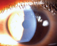
Figure 1. If striae limits BCVA, then immediate treatment is needed to prevent the striae from becoming embedded in the cornea. |
Infectious Keratitis
Fortunately, post-LASIK infectious keratitis is rare. The incidence rate after LASIK is low, reported to be between one in 1,000 and one in 5,000 procedures.1 Managing this complication begins before entering the surgical suite. As in the case of most postoperative ocular infections, the responsible organisms may derive from the lid flora. Important steps for infection prophylaxis include:
►Treat any pre-existing anterior or posterior blepharitis with tetracycline or a similar drug for 10 days prior to LASIK. Accompanied with warm compresses and lid scrubs, meibomian gland dysfunction may be controlled prior to surgery. The tetracylines have an antimicrobial effect and also improve the character of meibomian secretions. If treating a female patient, consider a prescription for an anti-fungal agent such as oral fluconazole 150 mg as a safety net to prevent candidal vaginitis.
►Surgical cap and povidone iodine application to lids and lashes prior to each case. Sequestration of lids and lashes with surgical drape.
►Involve a fourth-generation fluroquinolone in your regimen. Use one drop immediately after replacement of the LASIK flap and for 1 week after surgery. Pulsed dosing perioperatively or pretreatment 3 days preoperatively is not necessary in non-penetrating procedures due to the high activity and corneal penetration of these agents.
Over 50% of reported post-LASIK infectious keratitis cases are caused by atypical mycobacteria occurring between days 10 to 65.1 This condition is a challenge to treat. In cases that present earlier, the diagnosis is often delayed or confused with DLK. This not only causes appropriate treatment to be postponed but often compounds the problem as steroids, the primary treatment for DLK, aggravate the infection. The treatment is often plagued with multiple flap lifts and debridements that may ultimately culminate in flap amputation. A clue to the diagnosis of mycobacterial keratitis is consolidation of an infiltrate as opposed to the diffuse inflammatory infiltrate seen in DLK.
Although fortified topical amikacin and clarithromycin are the antibiotics of choice for mycobacterial keratitis, the fourth-generation fluoroquinolones have quickly gained popularity as a suitable alternative to these medications. They have good penetration, and even reach therapeutic concentrations in the aqueous humor.2 They demonstrate a broad spectrum of activity including gram-positive organisms as compared to earlier generations of fluoroquinolones.3
This improved coverage is an important consideration when realizing that these organisms represent over 90% of all ocular infections. A summary of the benefits and a safety profile of fourth-generation fluoroquinolones may be found in a recent article published in Current Opinion in Ophthalmology.4
|
|
|
Figure 2: Cyanoacrylate glue may be utilized for tissue adhesive in preventing epithelial cell migration. |
Striae
One of the more difficult obstacles when dealing with striae is determining their level of significance. Significant striae tend to decrease BCVA.
Insignificant striae occur at the level of the posterior corneal flap. These striae are generally not visually significant and resolve on their own by postop day 2 or 3. The cause of these striae is the area differential between the ablated stromal bed and the flap. They may be variable in their appearance depending on the amount of ablation performed.
A method to help minimize the occurrence of any kind of striae is to tap on the LASIK hinge once it is replaced, with minimal manipulation. Following irrigation of the flap stromal surfaces with balanced salt solution, the flap is repositioned. Then, the long-angled portion of the irrigating cannula is used on the hinge portion of the flap in a gentle tapping motion. The cannula is not rolled over the corneal surface because doing so may induce significant striae. Tapping the hinge with the cannula places two vectors on the eye which work to optimize proper flap positioning. The first downward vector increases the prolate shape of the cornea, stretching the ablated stromal bed so that it will better match the flap. The second horizontal vector distributes force across the flap to keep it in proper position.
Nasal hinges are also more prone to striae and sliding of the flap. They are mechanically less stable than superior hinges. When converting to the use of microkeratomes such as the Zyoptix XP (Bausch & Lomb, Rochester, N.Y.) with the capability to create nasal hinges — which also aids in mitigating dry eye postoperatively — use a bandage lens for 24 hours to maintain the stability of the flap.
Significant striae occur at the level of Bowman's layer (Figure 1). If the striae are central or found to be limiting BCVA, they must be treated immediately following surgery. Any delay may cause the striae to become permanently embedded in the superficial cornea.
In addition to lifting and repositioning the flap for pathologic striae, our technique employs a scrape-and-swell maneuver. First, an approximately central 3-mm by 4-mm epithelial defect is created. A Weck cell sponge is then soaked with sterile water and gently placed over the epithelial defect. Next, the cornea is irrigated with balanced salt solution to clear any epithelial cell remnants, after which the flap is lifted with a Sinsky hook and balanced salt solution on a cannula and then repositioned. Finally, a bandage contact lens is placed on the eye to help maintain proper flap alignment. Each step is designed to maximize rapid hydration of the superficial flap and break the irregular corneal configuration. In recalcitrant cases, the flap edge can also be sutured.
Epithelial Ingrowth
Epithelial cells may migrate into the interface postoperatively or clumps of these cells may be inadvertently introduced into the interface. The current incidence of epithelial ingrowth has been reported to be as low as 0.01%6 and as high as 61.1%,7 depending on a patient's risk profile. These risk factors include epithelial defects at the time of surgery, epithelial basement membrane dystrophy, hyperopic LASIK, flap instability and LASIK retreatments.
The diagnosis is often evidenced by the presence of whitish-grey opacification in the interface or by the appearance of clear translucent cysts often seen at the wound margin. Epithelial ingrowth may be confused with or seen in combination with DLK, as DLK is associated with signs of inflammation such as corneal haze or edema, conjunctival injection, foreign body sensation or pain and decreased vision.
Early intervention of this condition is key to successful management. Lifting of a preexisting LASIK flap places patients at an especially increased risk for epithelial ingrowth. These patients should routinely have a bandage contact lens placed and be examined with a slit lamp immediately postoperatively. A dry Weck-Cel sponge can be pressed over the contact lens to squeegee out any clumps of cells that may be present in the interface. We find this maneuver effective in removing epithelial clumps; in the vast majority of cases it alleviates the need to re-irrigate beneath the flap.
Routine care of these patients involves debridement of ingrowth at the leading edge of the flap. This is generally performed at the slit lamp, as the optics of many laser microscopes are not optimized for viewing epithelial irregularities and finding this edge may be difficult. The new Allegretto Wave (WaveLight, Erlangen, Germany) has an integrated slit lamp which could allow this procedure to be performed completely under the laser microscope. The vast majority of cases respond well to lifting the flap, scraping the bed and the posterior surface of the flap, and replacing the flap with a bandage contact lens. Suturing the flap may also help prevent cell migration.
Recently, there has been a great deal of discussion regarding tissue adhesive use for prevention of epithelial cell migration as an alternative to suturing. (Figure 2).
Taking the Right Steps
LASIK can be a life-enhancing procedure, and utilizing the aforementioned techniques will help to avoid or treat complications effectively and ensure positive outcomes for patients and surgeons alike.
Rafael Trespalacios, M.D., is chief resident of the University of South Carolina Department of Ophthalmology, and Richard Davis, M.D., is the chairman of the department.
|
Comprehensive
Ocular Surface Leads to Successful Refractive Surgery |
|
Refractive
patients achieve their best visual outcomes if they have had excellent care of the
ocular surface throughout their surgical experiences. Preoperative preparation,
meticulous intraoperative care, postoperative management and fastidious selection
of medications all help decrease the risks of complication and infection.
Prior to surgery, one of the most critical factors is confirmation of a normal tear film and corneal surface. Besides the normal use of artificial tears, appropriate management of pre-existing blepharitis is extremely important. In patients with dry eyes, the use of cyclosporine 0.05% (Restasis, Allergan) BID for 2 to 4 weeks preop often allows the cornea to heal better. By starting cyclosporine 0.05% early, the surface may normalize faster, particularly if there is an increase in dryness postop. The use of an NSAID such as ketorolac tromethamine 0.4% (Acular LS, Allergan) decreases inflammation and pain after surgery and improves patient symptoms as well. In surgery and following the procedure, the use of an antibiotic is critical. Gatifloxacin 0.5% (Zymar, Allergan) provides excellent broad-spectrum coverage as well as a rapid kill curve that decreases the risk of infection. It is biocompatible so that the ocular surface remains healthy and the postop healing process is as comfortable as possible. Following surgery, the use of the antibiotic for the first 4 to 5 days as well as close follow up is important. Patients who undergo surface ablation or who have increased sensitivity will benefit from the use of ketorolac 0.4% for pain control. It will also aid in the process of decreasing inflammation. Finally, patients should be restarted on cyclosporine 5 days postoperatively, or after they have stopped ketorolac 0.4% and gatifloxacin 0.3% treatment. This regimen will promote tear production and continue the process of preserving a healthy surface. In patients who have more significant symptoms from dry eyes, the use of punctal plugs may be beneficial as well. Refractive surgery outcomes continue to be refined and diligent selection of periop therapeutic regimens is an essential component of that process. Dr. RajPal, M.D., is principal of Cornea Consultants in McLean, Va. He can be e-mailed at cadcock@seeclearly.com. |
References
1. Karp CL, Tuli SS, Yoo SH, Vroman DT, Alfonso EC, Huang AH, Pflugfelder SC,Culbertson WW. Infectious keratitis after LASIK. Ophthalmology. 2003;110(3):503-510.
2. Solomon R, Donnenfeld ED, Perry HD, et al. Penetration of topically applied gatifloxacin 0.3%, moxifloxacin 0.5%, and ciprofloxacin 0.3% into the aqueous humor. Ophthalmology. 2005;112:466�469.
3. Callegan MC, Ramirez R, Kane ST, Cochran DC, Jensen H. Antibacterial activity of the fourth-generation fluoroquinolones gatifloxacin and moxifloxacin against ocular pathogens. Adv Ther. 2003;20:246�252.
4. Mah FS. Fourth-generation fluoroquinolones: new topical agents in the war on ocular bacterial infections. Curr Opin Ophthalmol. 2004;15:316�320.
5. Kang PC, Carnahan MA, Wathier M, Grinstaff MW, Kim T. Novel tissue adhesives to secure laser in situ keratomileusis flaps. Cataract Refract Surg. 2005;31(6):1208-1212.
6. Sun L, Liu G, Ren Y, Li J, Hao J, Liu X, Zhang Y. Efficacy and safety of LASIK in 10,052 eyes of 5081 myopic Chinese patients. J Refract Surg. 2005;21(5 Suppl):633-635.
7. Jun RM, Cristol SM, Kim MJ, Seo KY, Kim JB, Kim EK. Rates of epithelial ingrowth after LASIK for different excimer laser systems. J Refract Surg. 2005;21(3):276-280.


