feature
Attacking Cataract Surgery
Complications
Recent
advances create opportunities for both prevention and management.
BY STEVEN M. SILVERSTEIN, M.D.,
F.A.C.S.
Cataract surgical technique and technology is today nearly unrecognizable when compared to what was considered state-of-the-art just two decades ago. We follow studies of physician trends in each, as published by our organized societies, ophthalmology-related publications, and private marketing and research firms. From these sources, we learn that ours is a most rapidly evolving field. Ophthalmologists are never satisfied with maintaining the status quo, instead engaging in a thirsty crusade for ever-improving surgical performance, yielding yet better outcomes, with greater physician and patient expectations.
Physicians and companies are now working more closely than ever before to create new possibilities. We see exponential growth in both product development and application, as well as advances in operator technique, and though this equates to large potential net profits for industry and greater success for physicians, in the end, as it should be, it is the patient who wins, enjoying heretofore unparalleled measures of visual outcome.
The cost of research and development of new pharmaceuticals and devices is staggering, and it only takes one Vioxx (rofecoxib, Merck) scare to remind us that the process of technology advancement cannot be rushed, and, when a product fails to deliver, or worse, has deleterious consequences, the repercussions are swift and unforgiving. Ophthalmology has seen its share of such setbacks. Nevertheless, these are the necessary stepping stones toward progress, knowing now that we learn as much from our failures as from our successes.
Each success yields both greater patient acceptance of new technology, but also, greater expectation for both safety (diminished risk) and outcomes. We have seen this phenomenon very clearly in the field of laser vision correction, and we are seeing it manifest itself as well with both cataract and clear lensectomy patients.
Here, I'll discuss some of the more common complications associated with cataract surgery and how we are dealing with them with the tools and techniques of today.
The Risks Are Varied
Examining the risks and complications of cataract and refractive/presbyopic lens procedures provides great insight into how far we have traveled, and more important, what challenges we still face. Risk and complications arise from several sources, and will never be completely eradicated. Surgical learning curves for new physicians, the increasing age of the population (and thus, the greater need for surgical procedures), and the inability to control the patient's postoperative behavior, such as compliance with medication and maintaining proper hygiene, are examples of factors which will continue to make surgical risk a challenge. On the other hand, peri/intraoperative factors also create risk which may be impacted by our greater understanding of the pathogenesis of these complications.
|
|
|
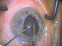
IFIS is characterized by iris billowing, prolapse to phaco and side-port incisions, and progressive miosis during phaco. |
Cystoid Macular Edema
Two decades ago, extracapsular cataract extraction was the predominant surgical technique preferred by most physicians. There was a higher incidence of vitreous loss and postoperative cystoid macular edema (CME), and theoretically, the greater surgical exposure time made the eye more susceptible to the development of endophthalmitis.
Surgical time was greatly lengthened with this approach, costing the healthcare system significant dollars in both resources and labor, and cost to the surgeon in terms of his/her optimal productivity. Currently, small incision, temporal, clear corneal, [no-stitch] surgery has minimized exposure to these factors, but by no means has eliminated them. Important studies are under way by each of the companies manufacturing nonsteroidal anti-inflammatory drugs, (NSAIDS) to determine whether they may make claims to the fact that these commonly used medications, when used in the peri-operative arena, are capable of lowering the risk of surgically induced CME. New imaging technologies, such as optical coherence tomography (OCT) which capture and document CME, are giving us greater insight into the layers of the retina implicated in this condition, as well as a noninvasive method of serially following the natural course of the disease, potentially leading to future medications which would be designed to specifically deliver medicine where it is needed most.
Endophthalmitis
Companies are also developing lens implants and IOL-delivery mechanisms to pave the way for sub-2 mm microsurgical instrumentation and technique. Smaller incisions suggest less risk for surface/tear organism contamination and a more secure, faster healing, astigmatically neutral wound. Certainly, since clear-corneal surgery became mainstream, it has raised the question of an increased risk of endophthalmitis, and studies by McDonnell et. al., demonstrating the ease of fluid entering the anterior chamber following a standard clear-corneal incision, have lent credibility to this argument1.
Our use and understanding of peri-operative antibiotics continues to broaden. The widespread use of fourth- generation fluoroquinalones such as Zymar (gatifloxacin, Allergan) and Vigamox (moxifloxacin, Alcon), with their remarkable MIC 90 aqueous concentrations following topical administration, has empowered us with unprecedented broad-spectrum coverage, combined with greater protection against bacterial resistance. Yet, we cannot let down our guard, as endophthalmitis continues to be a significant problem. Though many ophthalmologists continue to place an antibiotic such as vancomycin (generic) in the irrigating solution, and we all advocate the use of pre-operative topical antibiotics, still, nothing to date has been shown to reduce the incidence of postoperative infection more than the compulsive prep and drape of the eye and surgical field. The future addition of a sustained-release antibiotic/anti-inflammatory agent implanted in the eye at the conclusion of surgery is likely, and studies are currently ongoing which address the additive benefit of new methods of wound closure, such as the placement of a tissue adhesive along the wound edge.
Posterior Capsular Opacification
Posterior capsular opacification (PCO) has continued to elude our best efforts to date, though its incidence has fallen since the early days of standard PMMA IOLs. Though techniques have changed somewhat, such as the routine polishing of the posterior capsule prior to IOL insertion to remove and evacuate the largest concentration of crystalline lens epithelial cells, most of the progress in this area has come from work done on lens material and edge design.
Silicone and acrylic materials have been shown to decrease PCO, and several generations of lens-edge modifications have conclusively demonstrated an impressive halt in epithelial cell migration right up to, but not past the IOL perimeter, in a statistically significant number of cases. On the other hand, one study reported last year suggested that polishing the underside of the anterior capsule prior to implanting the IOL may actually increase PCO, and polishing was therefore not recommended. To what degree lens epithelial cells participate in the desired fibrosis of the anterior/posterior capsule/lens haptic complex is still uncertain, so the removal of all lens epithelial cells may produce a less stable IOL in the capsular bag, a potential problem for both potential dysphotopsia and especially for unwanted toric IOL rotation.
Astigmatism and Higher-Order Aberrations
Since the late 1970s, corneal refractive surgery has greatly furthered our understanding regarding the impact of aberrations upon the quality of our vision. Early on, our knowledge was significantly impaired by our inability to first capture, and ultimately analyze such information. We were limited to manual keratometry and placido disc-based instrumentation. Later, great strides were made with the first generations of automated topography, which provided reliable, reproducible numeric data combined with color-coded mapping, which truly shed light upon those components beyond the correction of myopia and hyperopia which influence how well we see. Advances in wavefront technology and its methods for interpretation have furthered our understanding regarding from where optical aberrations arise, (i.e., both the anterior and posterior surfaces of the cornea and lens).
This crucial knowledge has led to improved surgical techniques, such as customized placement and architecture of the wound and the routine addition of limbal relaxing incisions (LRIs). These advances have opened the door to a new generation of IOL design.
Five years ago, a biconvex square-edged foldable IOL was considered state-of-the-art. Today, the buzz surrounds available and future aspheric optic technology, with a choice of hydrophobic or hydrophilic material, and an apodized diffractive, or refractive multifocal or pseudo-accommodating IOL, or a combination of each.
This smorgasbord of lens options is analogous to ordering at Starbucks, where we no longer simply order a cup of regular or decaf. Instead, we must now select the coffee beans' country of origin, and then dress it up or down with flavor and calories. No wonder patients are confused. In the future, lenses will be available for custom order, with the patient's unique wavefront pattern and refractive preferences pre-lathed into the optic.
An Expanding Circle of Change
The changes in lens design do not stop at affecting patient outcomes and expectations. They have also made a precedent-setting impact upon society, where, for the first time, IOL pricing has climbed to nearly $900, thus paving the way for a radical departure in federal government policy, allowing the opportunity to balance bill for access to advanced technology. Even the sociology and culture of where ophthalmologists perform these surgeries has changed dramatically, with nearly every state in the country seeing an explosion of outpatient, often physician-owned, ambulatory surgery centers. As we have learned once again from the corneal refractive marketplace, with advanced technology and higher expectations comes the potential for greater patient dissatisfaction, and an increase in medical-legal exposure. At the very least, discussing the options available to patients today has significantly increased the doctor-patient chair time, the need to hire additional surgical counseling personnel, or both.
With advanced lens technology, in addition to newer methods of phacoemulsification (venturi vs. peristaltic, laser vs. ultrasound, bimanual vs. coaxial microincisional), comes the development of products which increase the accuracy and the safety of lens surgery. More sensitive methods of biometry and its associated IOL-calculating formulas, especially in eyes that have previously undergone kerato-refractive surgery, are being developed. New generations of viscoelastic agents (OVDs) combining optimal cohesive and dispersive characteristics further our understanding of tissue protection and manipulation as well as IOL delivery, for many of the products that are introduced into the eye through an insertion device.
Interestingly, some of the challenges in cataract and refractive/presbyopic lensectomy arise from sources out of our control. Prior to the common practice of using a topical anesthetic agent, surgeons typically discontinued the use of blood thinners such as aspirin and Coumadin (warfarin, Bristol-Myers Squibb) to reduce the likelihood of a retrobulbar hemorrhage associated with the eye block. This not only placed the patient at significant systemic risk, but such peri-ocular (lids and face) or retrobulbar hemorrhage was still often seen.
Today, thanks to research by David Chang, M.D. and others, Flomax, (tamsulosin, Boehringer-Ingelheim), an alpha 1a adrenergic antagonist used to facilitate urine flow in patients with benign prostatic hypertrophy, has been revealed as a new nemesis, causing intraoperative floppy iris syndrome (IFIS), or poorly dilating pupils. Discontinuing Flomax use preoperatively typically has no effect and surgeons must often carefully work around a prolapsed iris or a small, distorted pupil.
Prepare for More Change
We have progressed greatly in a remarkably short period of time. The evolution of lens-based procedures affects each of us, first as physicians, and ultimately as patients. Driving forces such as industry, patient demand, economic considerations, an aging population, governmental influences, and ultimately, our innate scientific curiosity, will continue to push us further along the continuum ever faster. I look forward to seeing how far we have traveled when I ultimately step aside at the end of my career, and observe and speculate what will be possible as we continue to work and learn together.
Steven M. Silverstein, M.D., F.A.C.S., is a partner in Silverstein Eye Centers, P.C., in Kansas City, Mo. He is also clinical professor of ophthalmology at the University of Missouri Kansas City Medical School and clinical professor of ophthalmology at the University of Health Sciences. He can be reached via e-mail at ssilverstein@silversteineyecenters.com
Reference
1. Ingress of India Ink into the Anterior Chamber Through Sutureless Clear Corneal Cataract Wounds: Taban M, Sarayba MA, Ignacio TS, Behrens A, McDonnell PJ: Arch Ophthalmology, 2005 May; 123(5):643-8.


