Surgical Pearls for the Femtosecond Laser
How
we create successful IntraLase flaps.
WILLIAM
W. CULBERTSON, M.D.
We have used the IntraLase femtosecond laser (IntraLase, Irvine, Calif.,) to create 99% of my LASIK flaps for the last 2 years. We had acquired this technology after considerable investigation and due diligence, with the dual goals of reducing the potential for serious flap-related complications and improving the consistency of flap dimensions. Following a short learning period, we think that we have realized our original objectives and have been pleasantly surprised by some unanticipated benefits such as enhanced accuracy of correction and good early postoperative visual acuities.
|
|
|
|
Figure 1. Dry stromal bed created by the IntraLase
laser. |
|
How IntraLase Works
As currently designed, the IntraLase near-infrared laser creates a LASIK flap quite differently from a blade microkeratome. Short 700 femtosecond pulses, 2.3 μm in diameter, are placed in an overlapping pattern in the cornea. A lamellar plane and a vertical edge cut are thus created in the corneal stroma to fashion the flap. Although the cornea is applanated, there is no movement of a microkeratome plate across the corneal epithelium. The lack of movement minimizes the chances that sheering stress on the epithelium will cause an epithelial abrasion or slide. Water and debris are not pulled into the interface because the IntraLase procedure is performed under dry conditions without any need for lubrication. Thus, the flap beds retain their natural consistent hydration and the surgeon does not have to contend with irregular hydration of the flap causing unpredictable ablation effects (Figure 1).
With the IntraLase, the dimensions and orientation of the flap are custom-designed by the surgeon using computer software. The precise diameter, thickness, centration, hinge angular width, hinge orientation and edge verticality of the flap are selected by the surgeon based on the most desirable parameters for the individual eye.
|
|
|
|
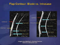
Figure 2. Artemis II views of the cross-sectional
profile of the meniscus flap created by a blade microkeratome compared to planar
shaped IntraLase flap. |
In contrast, the dimensions of a flap cut with a blade microkeratome are less consistent and are dependent on certain features of the cornea such as keratometry and thickness. Because the flap dimensions are predictable with the IntraLase, I feel more comfortable performing LASIK on thinner, flatter and steeper corneas than I would with a blade microkeratome. For instance, if I need a 100-μm flap in a myopic eye requiring a deeper ablation, I can be reasonably assured that I can create a flap from about 90 to 110 μm. Similarly, I can make a large 9.2-mm diameter flap in a 40 D flat cornea for a hyperopic treatment without fear of a free cap. Steep corneas with keratometry above 46 D are not vulnerable to buttonholes using the IntraLase, so I am able to extend the range of treatable corneas to K-readings of 47 D through 50 D if there is no other contraindication to LASIK.
I usually orient the flap hinge superiorly, but if I want to place it on the nasal side to optimize flap innervation or to avoid a pterygium, I can just enter "nasal" into the software prior to cutting the flap.
Flap Consistency
Another advantage of using the IntraLase is that the thickness is the same across the entire flap (Figure 2). I believe that this feature is responsible for minimizing flap-induced refractive effects and higher-order aberrations such as coma and trefoil. Also, the edge of the flap can be cut more vertically (Figure 3). I use a 70Þ to 80Þ side cut angle so that the flap fits in the bed like a manhole cover and is less likely to be displaced by minor trauma during the early postoperative period. This vertical edge cut may be responsible for my observation of the absence of clinically significant epithelial ingrowth (more than 0.25 mm) either after primary LASIK or enhancements. We take advantage of this effect in enhancements of previous blade microkeratome-created flaps. I use the IntraLase to make a 70Þ vertical edge cut of only 0.3 mm inside the old flap edge and then lift the flap and perform the retreatment. This maneuver tends to decrease the risk of epithelial ingrowth following enhancements and allows the edge to heal more firmly than with a beveled-blade microkeratome, decreasing the risk of late traumatic flap displacement.
|
|
|
|
Figure 3.
The thinner vertical edge of the IntraLase bed compared to the thicker beveled edge
of a bed created with a blade microkeratome. |
|
I am able to center the flap on the pupil by placing the suction-docking patient interface concentric with the pupil. Once docked to the IntraLase laser, the location/centration of the flap can be more finely adjusted with the computer software (Figure 4). With the adjustment, the excimer laser ablation is concentric with both the pupil and the center of the stromal bed. I believe this improved bed/flap centration results in better objective and subjective visual outcomes.
The IntraLase may have more limited usefulness for making flaps in eyes that have had previous incisions such as in PK or RK. In these cases, care must be taken to avoid dehiscence of the incision when lifting the flap. I prefer to use a blade microkeratome or perform surface ablation if I feel that the incisions are not tightly healed.
Limited Side Effects
We have not experienced any cases of free caps, buttonholes, incomplete flaps, incomplete free flaps, abrasions or infection during our first 2 years of using the IntraLase for LASIK. Although our incidence of overall serious flap complications has been markedly reduced while using the IntraLase for LASIK flaps in over 2,000 eyes, we have noted an interesting set of new side effects that we had not encountered with the blade microkeratome.
For example, in two eyes, the flap could not be lifted because of inadequate interface dissection of uncertain cause. One of these eyes underwent immediate surface ablation and the other had successful LASIK 3 months later with a blade microkeratome. In another case, a beginning IntraLase user inadvertently passed the end of the lifting spatula through the edge of the flap and replaced it without consequence. Surface ablation was carried out later.
|
|
|
|
Figure 4. The IntraLase software can be employed
to center precisely the flap concentric with the pupil. |
We have experienced a few cases of self-limited non-diffuse interface keratitis that responded to a 5-to-10 day course of topical steroids. During our first month of IntraLase use, we had one case of severe diffuse lamellar keratitis-like inflammation occurring at the edge of the flap. It was successfully treated with topical steroids with a final 20/20 visual result. We felt that the cause of this episode was too high of a side cut energy setting.
Another side effect that we have seen with the IntraLase has been transient photophobia occurring approximately 2 weeks postoperatively. This syndrome, which has been termed good acuity photophobia syndrome (GAPS), is invariably benign and is treated with a short course of weak topical steroids.
LASIK with Confidence
The combination of a dependable IntraLase flap, wavefront-guided laser ablation, with tracking and iris registration/centration has resulted in a more predictable, safer LASIK procedure. Happily, our ophthalmologists and technical staff at the Bascom Palmer Eye Institute Refractive Surgery Center feel a sense of renewed comfort recommending and performing LASIK for eligible patients.
William W. Culbertson, M.D., is professor of Ophthalmology and director of Refractive Surgery at Bascom Palmer Eye Institute, University of Miami. He can be reached via e-mail at wculbertson@med.miami.edu.


