instrument insider
The Sabet Safety Net
By Sina J. Sabet, M.D.
The dropped lens during cataract extraction remains one of the most dreaded complications that cataract surgeons face. In this age of improving technique, equipment and fluidics, its occurrence still haunts experienced surgeons, as well as ophthalmology residents struggling through the learning curve of phacoemulsification. Numerous ways of handling this difficult complication have been proposed, none of which has yet been proven to be completely satisfactory. One answer is the posterior-assisted levitation (PAL) technique described by Richard Packard, MD, and the late Charles Kelman, MD.1,2 This article introduces a new instrument, the Sabet Safety Net, which attempts to build on the PAL technique with a safer and more effective instrument, allowing the handling of this complication at the time of the primary procedure.
|

|
|
|
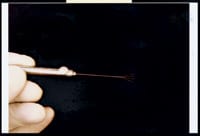
Figure 1. The Safety Net in a side profile in a closed position. |
|

|
|
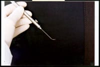
| Figure 2. The Safety Net in a superior profile with the button depressed and the net in the open position. |
The Problem
In the original PAL technique, a second instrument, often a cyclodialysis spatula, is inserted through a paracentesis incision to bring the dropping lens fragment up into the anterior chamber and to support it for further phacoemulsification (a pars plana incision may also be used according to Dr. Kelman). Alternatively, the fragment can then be expressed whole through an enlarged extracapsular incision. The problem with this procedure is that any instrument narrow enough to fit through a small paracentesis incision will not provide enough stability to reliably bring the lens up into the anterior chamber or to adequately support it for vitrectomy or phacoemulsification. The fragment easily wobbles off the second instrument. This is especially true if there are multiple fragments or the fragment is relatively small. Even if the lens fragment is successfully brought up into the anterior chamber, good support for continued vitrectomy/phacoemulsification will often require the placement of a Sheets glide. The Sheets glide cannot be wider than the approximately 3 mm clear corneal or scleral tunnel incision and can place significant traction on any vitreous that may be present.
The Solution
The Sabet Safety Net is designed to address these shortcomings. Its bore fits smoothly through a paracentesis incision (Figure 1). If desired or necessary, a pars plana incision can also be used. When the button is depressed, four malleable metallic wires exit through the bore and fan out, providing an area of support measuring approximately 6 mm at its widest point (Figure 2). This provides stability for the lens fragments and allows them to be brought up from the vitreous cavity into the anterior chamber or iris plane. It also allows vitrectomy and phacoemusification to continue without any need to enlarge the incision.
When viewed from the side, it can be seen that there is a relatively steep angle formed by the metallic wire and the bore of the instrument (Figure 3). This is to allow the bore to be placed vertically into the vitreous cavity, but to keep the wires horizontally behind the lens fragment when they are opened. The use of only four thin wires rather than mesh or an elastic net is to minimize traction on the vitreous in the course of the initial intraocular manipulations, when the lens fragment may be completely enmeshed in the vitreous gel. This rather loose net design may allow small fragments to drop back, but these would then resorb spontaneously and most likely not require additional surgery.
|

|
|
|
Figure 3. The Safety Net in side profile in an open position. |
|

|
|
| Figure 4. A higher magnification view of the tips of the net wires. |
Another design feature can be seen in a higher magnification view of the tip of the wire mesh as seen from the side (Figure 4). The tips are bent forward to provide traction on the lens fragment and to keep them from becoming displaced anteriorly. The bowled shape to the net also allows the fragments to remain more stable inside of it. If there is any inadvertent touching of the retina during the procedure (an unlikely event if the handle is held vertically), the bend in the wires ensures that the sharper tips do not induce any tears.
Once the fragment is secure, vitrectomy can then be done to free the fragment from the vitreous. If the lens is soft, the vitrector can be used to remove the lens material as well. If dealing with a hard fragment, however, phacoemulification can be performed to remove it. Depending on the circumstances, consideration can also be given to enlarging the incision and expressing the fragment. With the fragment sitting securely on the Safety Net, the surgeon has the luxury of time in deciding on the best method of attack.
Critique
One criticism that can be made of this instrument (and has been made of the PAL technique as well) is the higher risk of inducing retinal tears and detachments through extensive intraocular manipulations. Better to let the fragment drop and have a vitreoretinal surgeon remove it later than to risk a retinal tear, critics argue.
There are several responses to this. First, the risk of a retinal tear is already high when the capsule is ruptured. Studies would have to be done to determine if using the Safety Net would indeed increase the risk beyond the initial risk associated with the capsule rupture. Some initial studies have already begun to show favorable outcomes with the PAL technique.3 Second, even if there is an increased risk, this risk would need to be weighed against the alternative risk of a second procedure involving pars plana vitrectomy and lensectomy. Third, this risk would have to be weighed against the patient and family's response to a dropped lens, the specter of having to go back to the operating room with another surgeon for yet another major surgical procedure, and the anxiety, pain and discomfort associated with the difficult to control inflammation and elevated IOP. It is easier to explain to the patient and family that there was a broken capsule during the case, but that the cataract was removed in its entirety and the IOL was inserted. It could then be explained to them that the patient will be followed closely in the office because of a potential increased risk of retinal tear; something that if caught early can be fixed with an office laser procedure. It would be unlikely to require another operating room procedure for repair of a full-blown detachment. The patient can then be monitored postoperatively by either the primary surgeon or a retina specialist. Finally, many cataract surgeons, despite the valid objections raised by critics, are even now fairly aggressive in chasing lens fragments once they begin to drop for the above reasons. The Safety Net simply provides safer instrumentation to allow them to do this.
Sina Sabet, M.D. is in private practice in Alexandria, Va. Dr. Sabet is active in teaching ophthalmic surgery and pathology at Georgetown University.
References
1. Packard RBS, Kinnear FC. Manual of Cataract and Intraocular Lens Surgery. Edinburgh, Churchill Livingstone, 1991;47.
2. Kelman C D. Posterior Capsule Rupture: PAL Technique. Video J Cataract Refract Surg. 1996;12:no 2.
3. Lifshitz T, Levy J. Posterior assisted levitation: Long term follow-up data. J Cataract Refract Surg. 2005;31:499-502.


