Epi-LASIK: the Procedure, Pain
Management and Patient Results
Providing a better
understanding of epi-LASIK surgery and its aftermath.
BY MARK VOLPICELLI, M.D., WITH CONTRIBUTIONS BY WILLIAM TRATTLER, M.D.
As a surgeon who has performed laser-based refractive surgery on thousands of patients over the past 10 years including family members and my ophthalmology partner, I would like to provide insight into some recent changes and advancements in epi-LASIK technology. Epi-LASIK is a predictable technology; most obviously, because the corneal stroma is not cut, many of the complications associated with laser vision correction are avoided. In addition, epithelial separators travel across the eye more slowly than microkeratomes, providing better control over the separation. Epi-LASIK allows the surgeon to create a healthy epithelial sheet easily and consistently.
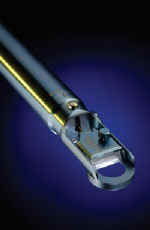
The development of epikeratomes with non-cutting or non-sharp separators, such as the Norwood EyeCare Epikeratome (Duluth, Ga.)(Figure 1), has helped minimize the stromal incursion rate (the rate at which the separator dives too deeply and removes a small anterior stromal divot). With any sharp separator, there is a risk of cutting into Bowman's layer, leaving the natural cleavage plane with all the associated problems. The Epikeratome is user-friendly � it is light and ergonomic. Because it is similar to my preferred microkeratome, the Nidek MK2000 (Fremont, Calif.), I did not need to develop a new skill set.
The design and placement of the suction ports on the Epikeratome is important for acquiring vacuum effectively and keeping the separator on the natural cleavage plane in a variety of eyes, including deep-set or narrow eyes. With the Epikeratome's vacuum head features, such as the advanced castellations/fenestrations, I have never broken suction, and have had no problem creating an adequately sized epithelial sheet.
The two options for suction ring size (9 mm or 10 mm) enable me to work on eyes of various size as well as those with smaller orbital anatomy. The smaller ring also completely avoids the limbal stem cells, thus reducing the chance of involving epithelial stem cells at the limbus and reducing healing time.
Some of the available epi-LASIK systems use an applanator to flatten the epithelium in front of the separator as it passes over the cornea. In the case of the Epikeratome from Norwood EyeCare, the separator's design incorporates a posterior applanation platform to flatten the cornea as it advances. This unique design feature serves to both applanate the cornea and to minimally increase vacuum.
Introducing Epi-LASIK Into Practice
I currently use epi-LASIK for patients who do not want a surgical flap on their cornea, who want minimal risk and desire no cutting, as well as patients who might otherwise not be good candidates for LASIK or an IntraLase procedure based on age, corneal topography, pachymetry and/or vocational/extracurricular activities such as participation in martial arts or contact sports. In addition, epi-LASIK is the perfect niche for a patient with flat, steep or thin corneas; for an older patient whose epithelium has the potential for an epithelial slide with LASIK; and for a patient with apparent basement-membrane dystrophy. It is the best option for patients like these, while at the same time a valid option for a wide range of patients.
Some types of patients should not undergo this procedure, especially those who have had previous refractive surgery. If Bowman's layer is gone because of PRK or LASEK, you may not obtain the correct plane for the epi-LASIK procedure. With post-LASIK patients, you run the risk of creating a buttonhole in the epithelial sheet. Another group of patients who should not undergo epi-LASIK are those who have undergone a corneal transplant, for similar reasons.
|
|
|
|
Figure 1. Norwood Epikeratome Handpiece |
|

|
|
|
Figure 2.
Decrease in percentage of eyes with postop pain over 24 hours (n= 163 eyes) (Courtesy of Vikentia Katsanevaki, MD.) |
|

|
|
| Figure 3. Average Snellen UCVA (n = 22 eyes) |
The Bed-Sheet Analogy
I use the analogy of a bed-sheet and a mattress: epi-LASIK only lifts the sheet and leaves the underlying mattress intact, while the flap-making techniques cut into the upper part of the mattress and lift both the bed-sheet and upper mattress together as a unit. William Trattler, M.D., at the Center For Excellence In Eye Care in Miami, also performs epi-LASIK with the Epikeratome. He describes epi-LASIK to patients as "being like a snowplow that separates and slides the epithelium off like a layer of snow, which is replaced intact."
No matter how you describe epi-LASIK, the surgeon must prepare the patient to accept a slower visual rehabilitation than LASIK patients. Their vision may initially be fuzzy and/or blurred for the first several days because their epithelial cells will not be perfectly aligned as in an unoperated eye and the eye takes longer to heal. Most patients recover in 4 to 5 days but it is important that the surgeon not over-promise rapid visual acuity recovery and have patients experience disappointment as a result.
Pain Control
The first few days, of course, are not comparable with LASIK with regard to comfort, but I am seeing much less pain than with PRK. I use preoperative and postoperative NSAIDs, along with dilute tetracaine and Vicodin (hydrocodene/acetominphen, Abbott Laboratories) as "escape medications" for the first 48 to 72 hours if needed. Using cold balanced salt solution before and after for 30 seconds also helps quell the inflammatory response and helps with pain control. Fewer than 20% of my patients have experienced significant discomfort; analysis is still in progress on my patients' objective comfort data, including pain scores and their use of pain medication during the first 72 hours postoperatively.
Dr. Trattler uses Celebrex 200 mg/day (celecoxib capsules, Pfizer), starting 1 day preop and extending to 5 days postop, and also provides dilute tetracaine, which patients use up to every 1 hour as needed (most patients use the dilute tetracaine just a few times). He has also found that using extremely cold balanced salt solution both at the beginning of the surgery and directly after the ablation can help reduce postoperative pain. Dr. Trattler also uses a special regimen for dry eye patients, whom he pretreats before surgery to achieve a better outcome. He pretreats with Restasis b.i.d. (cyclosporine ophthalmic emulsion, Allergan) and Soothe t.i.d. to q.i.d. (metastable emulsion, Alimera Sciences); placement of punctal plugs; and use of preservative-free tears (Systane [lubricant eye drops, Alcon], Refresh [lubricant eye drops, Allergan]) and Refresh liquigel during the epithelial healing stage. Dr. Trattler's patients are also given an "emergency bottle," a 1-cc bottle of preservative-free 0.5% tetracaine, with instructions to use it only for the incidence of severe pain. He reports that only a small percentage of epi-LASIK patients require the full strength tetracaine, and typically one or two drops are all that is needed.
Data from Ioannis Pallikaris, M.D., in Crete show that his patients experience less pain and recover faster with epi-LASIK than with PRK. In a recent analysis of pain scores for 163 eyes treated with epi-LASIK for moderate myopia, at 2 hours, only 12% reported pain higher than 1.0 ("discomfort") and that percentage dropped to 2% at 8 hours (Figure 2). In a similar study of 92 eyes conducted by Efekan Coskunseven, M.D., 80% of patients reported no pain or major discomfort.1
The Role of the Bandage Contact Lens
Our protocol currently calls for a Soflens 66 (Bausch & Lomb, Rochester, N.Y.) and it has worked well to prevent medication and debris accumulation, as well as for improved comfort. Dr. Trattler reports his patients have been extremely comfortable with the Acuvue Oasys (Vitstakon, Jacksonville, Fla.) contact lens, a new contact lens designed for dry eyes with a high dK (more oxygen permeability to help the healing).
Of course, in a sense, epi-LASIK by definition leaves a natural bandage on the eye instead of an open wound. The separated sheet protects the healing surface for the first few postoperative days to facilitate better and faster healing. Leaving the epithelium on the eye, including the basement membrane, blocks many of the inflammatory pathways that stimulate pain. Epi-LASIK accurately separates the epithelial sheet above Bowman's layer but below the basement membrane. With an intact basement layer, fewer inflammatory signals are sent to the keratocytes.
Visual Recovery
We have just finished analysis of the 3-month data for the center's first 22 of 30 epi-LASIK eyes, as part of Norwood EyeCare's U.S. Prospective Study. Patients' average age was 38, ranging from 29 to 67; 45% were male and 55% female. Average preoperative corneal pachymetry was 527 μm, ranging from 469 μm to 605 μm. All patients were treated using the VISX Custom Ablation platform (AMO, Inc., Santa Ana, Calif.).
The results confirmed what we know about epi-LASIK; visual recovery is slower than LASIK � though more rapid than PRK or LASEK cases � and improves dramatically by 1 month, with further improvement at 3 months. Average Snellen UCVA was 20/53 on postop day 1, and 20/43 on day 7 (see Figure 3). However, at 1-month postop, average UCVA was 20/20, and at 3 months the average was 20/17. This compares favorably to typical 3-month UCVA results with custom LASIK.
Average preop manifest for these patients was -2.56 D, ranging from -0.25 D to -5.75 D. At three months postop, average manifest was +0.05 D, ranging from +0.05 D to -0.25 D. At 3 months, average spherical equivalent was 64% at plano, and 91% were between -0.25 D and +0.25 D of plano.
Results for corneal haze also confirm that while the healing of the epithelium after epi-LASIK creates the appearance of the epithelial remodeling that occurs after PRK or LASEK, there is usually scant to no haze by the end of the first month and virtually no haze by the third month. At day 1 postop, haze measurement (on a scale of 0-4) indicated a value of 1.5, which decreased to 0.5 at day 4. At 1 month, haze measurement indicated a value of 0.7, but it decreased to 0 at 3 months.
Dr. Trattler told us he has seen no haze to this point. His epi-LASIK procedure for higher risk eyes, starting with 6 D to 7 D of myopia, includes low-dose (0.02%) mitomycin-C for 12 seconds to prevent haze.
Handling Less Than Perfect Results and Retreatments
Retreatments have not been necessary to date in our study patients. However, I would probably enhance with conventional alcohol-assisted LASEK and/or PRK to be certain there was no stromal intrusion in these "non-virgin eyes."
Epi-LASIK allows us to avoid many of the complications that can occur with LASIK. Additionally, epi-LASIK allows for potentially faster epithelial healing and less discomfort compared to other forms of surface ablation. It is safe, and ultimately, patients do extremely well. The safety issue is paramount, because one of the main causes of patients' rejection of laser vision correction is fear of complications and potential side effects such as glare and night vision difficulties.
Reference
1. Coskunseven, E. Epi-LASIK for low myopia: 1-year results in 92 eyes. Paper presented at the ASCRS/ASOA 2005 Symposium on Cataract, IOL and Refractive Surgery; April 16, 2005; Washington, D.C.
Mark Volpicelli, M.D., is in private group practice at Peninsula Laser Eye Medical Group in Mountain View, Calif., one of the three study sites for Norwood EyeCare's U.S. Prospective Study. He is on the clinical faculty in the Department of Ophthalmology at Stanford University. He states that he holds no financial interest in any product or company mentioned herein. Dr. Volpicelli may be reached at (650) 961-2585; volpeyes@aol.com. William Trattler, M.D., is the cornea specialist for the Center For Excellence In Eye Care in Miami, Fla. and is a volunteer assistant professor at Bascom Palmer Eye Institute.


