spotlight on technology & technique
An Emerging Technology from Optos
Optos' P200MA fluorescein angiography produces images of approximately 200° of the retina.
By Leslie Goldberg, Assistant Editor
Optos' P200MA (Optos, Marlborough, Mass.) produces ultra-widefield, dynamic high-resolution angiographic images of the retina. This new technology builds on the platform of the established P200 instrument, a scanning laser ophthalmoscope that produces the optomap retinal exam — a 4MPixel image of the choroid and retina. The addition of the P200MA enables doctors to get the same ultra-widefield view of the retina, but with angiographic capability.
Optos' technology uses low-powered red and green lasers to scan approximately a 200° interior angle of the retina and converts the reflected light into the digital optomap image. The addition of a blue, 488nm laser to this platform allows ophthalmologists to perform fluorescein angiography (FA) of the same ultra-widefield view, creating a stack of sequential images called optomap fa. The optomap fa dynamic ultra-widefield angiographic study is a more practical and valuable approach than montaged narrow field-of-view images that only provide a composite from different parts of the retina taken at different points during the circulation of fluorescein says Optos.
|

|
|
|
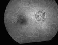
This image shows a regular angiogram, displaying only the central pole. |
|

|
|
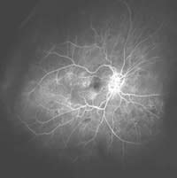
| An optomap fa dynamic ultra-widefield angiography image in its full 200° field of view. | |

|
|
| This is the same image zoomed in on or about the same area as the traditional angiogram.
IMAGES COURTESY OF IVAN� SUNER, M.D. |
The Addition of FA
Optos has developed the P200MA instrument as a retinal imaging platform capable of producing three image types that can be selected for different situations:
► the standard optomap retinal exam – for screening and wellness examinations;
► the optomap plus medical retinal exam – enhanced capabilities (resolution, eye steering and zoom) used to assist diagnosis or for other medically necessary procedures; and
► the optomap fa dynamic ultra-widefield angiography – for high resolution angiographic retinal analysis.
Physician Feedback
Clinical trials performed recently on evaluation devices have created considerable interest from retinal specialists. "The ability to capture the peripheral characteristics of patients will most likely revolutionize angiography," says Thomas Friberg, M.D., professor of ophthalmology and bioengineering and chief of the retina service at the University of Pittsburgh School of Medicine.
"This new technology is extremely helpful when handling patients who may have diabetic retinopathy," says Dr. Friberg. "Pilot studies have shown that many patients who have diabetic retinopathy display signs far in the periphery of the retina. We were unable to see these changes prior to this advanced FA technology."
"Prior to Optos' new technology, we were only able to capture multiple 30°-45° images of the retina. Viewing out to the edges of the retina greatly increases our chances of catching capillary drop-out and neovascularization that previously went unseen," says Roger Novack, M.D., Ph.d., a vitreoretinal specialist at Retina-Vitreous Associates Medical Group in Los Angeles, Calif. "I believe that the [P200MA] will eventually make other angiographic techniques obsolete. The ability to take pictures quickly with a panoramic view, zoom in to a point of interest and capture important findings using Optos' proprietary screening software program is advantageous."
The clinical trials were a multi-center pilot study, and indicated a statistically significant increase in the mean retinal image area obtained compared to conventional digital FA imaging systems, with 45% more retina being imaged. Results also indicated that significantly more diabetic pathology (neovascularization and ischemia) was identified by the Optos instrument. The ability to better visualize areas of peripheral ischemia will likely lead to earlier identification of eyes at higher risk of disease progression and earlier intervention says Optos.
Additional features of the P200MA system include:
► An image recognition system that facilitates patient positioning
► DICOM compatibility
► Patient-gaze angle control
► Proprietary software that assists doctors in correct documentation for reimbursement claims
► A patient interface designed for comfort and stability.
Optos' focus
Ease of use has been a primary focus for Optos. All Optos instruments are designed to image through a minimum of a 2-mm aperture and therefore, the decision to dilate the patient can be made by the physician purely on medical grounds alone.
High-quality images can also be obtained through opacities, such as moderate cataracts, and all images produced by the Optos instruments provide a permanent medical record on an image server that can be integrated to leading EMR software packages. The digital images can be enlarged, manipulated, stored and readily transmitted to other healthcare providers through the proprietary software.
Contact Information
For more information on Optos' P200MA, please call 1-866-OPTOMAP.


