marketplace
Focus on Fundus Cameras

CANON CF-60DSI
Canon Medical Systemsa division of Canon U.S.A., Inc.
Retinal examinations can be conducted with greater efficiency with Canon's CF-60DSi Digital Fundus Camera, an all-digital system offering PC-based operation and study management, high digital image quality, immediate image viewing and DICOM (Digital Imaging and Communication in Medicine) networking capabilities.
The company's Canon fundus camera optics provide 60° field-angle coverage in order to capture a larger area of the retina with each image. The Canon CF-60DSi also offers close-ups to support diagnostic requirements in ophthalmology and software to manage workflow.
The Canon CF-60DSi Control Software has functions for image capture, study worklist management, image transfer, printing, QA viewing and temporary storage. For each captured image, the software assembles the image,
patient/study data, camera information and timer data into a comprehensive medical data file suited for diverse image management systems.
Canon Medical Systems
Phone: (800) 970-7227
www.usa.canon.com/eye-care

NONMYD μ-D NON-MYDRIATIC DIGITAL FUNDUS CAMERA
Kowa
Kowa's Nonmyd μ-D non-mydriatic digital fundus camera is a compact, all-in-one unit that takes advantage of high-resolution digital imaging, a user-friendly interface, and increased speed, accuracy and ease of use. The camera's controls are positioned within easy reach, allowing for rapid screening without the need to search for switches.
The Kowa designed digital optics and built-in megapixel digital camera provides the imaging power for the NonMyd μ-D Non-mydriatic fundus cameras superior image quality. Kowa states that the convenient VK-2 imaging software allows it to
interface and communicate with virtually any office network PC with plug-and-play ease. Two picture angles are available when using the camera: 45° and 20°, and a 5.6 inch LCD monitor provide the user with an easy alignment and focusing interface.
Kowa
Phone: (800) 966-5892
www.kowa.com

NM-200D
NIDEK
NIDEK delivers performance, compact design and portability with its digital handheld non-mydriatic fundus camera, the NM-200D. The NM-200D features a lightweight body with user-friendly color touch screen display. The clear 10.4-inch LCD screen offers ease of operation and functionality for image capture, processing, editing, zooming and transmission of image data to the compact flash card. Through USB interface, you can quickly transmit the NM-200D's high-resolution, 1.5 mega pixels to NAVIS or other devices. You can also correct and display its high-quality images, stored in .tiff format, with other applications. In addition, the NM-200D's high-resolution CCD camera requires a much lower flash intensity than 35-mm or Polaroid film type fundus cameras, translating into increased comfort for patients.
Nidek
Phone: (800) 725-4562
www.Nidek.com

TRC-50EX AND TRC-50IX FUNDUS CAMERAS
Topcon
Topcon offers a full line-up of retinal cameras including the TRC-50EX and TRC-50IX Mydriatic Fundus Cameras, featuring expanded flash settings, alignment dots for new users and excellent image quality. A variety of resolutions and appli
cations are available, including the new 11MP FA/Color System or 3 MP ICG System. All Topcon cameras integrate seamlessly with Topcon's IMAGEnet software to create a digital retinal capture system, allowing for easy image comparison/manipulation, image archive/transmission and an array of quantitative analysis tools and networking capabilities.
Topcon
Phone: (800) 223-1130
www.topcon.com

CARL ZEISS MEDITEC'S
FF450 plus Professional Fundus Camera
Carl Zeiss Meditec's FF450 plus fundus camera provides three field angles - 50°, 30° and the smaller 20° field for precise detail of the optic nerve and macula. The FF450 plus delivers images for the diagnosis of retinal diseases with an extremely high level of accuracy using "Zeiss telecentric optics." These optics guarantee precise retinal measurement, while providing a light yield with minimal
exposure to the patient. With capture mode options of color, red, red-free, blue, fluorescein and ICG, the camera's versatility makes it practical for all retinal imaging applications. The FF450plus ensures fast, easy operation with motorized filters and advanced exposure adjustment capabilities with extensive default settings for every capture mode. Extensive viewing, archiving and data management capabilities are standard with every FF450plus when integrated with the Zeiss VISUPAC Digital Imaging System.
Carl Zeiss Meditec
Phone: (800) 342-9821
www.meditec.zeiss.com

THE ARIS
Visual Pathways, Inc.
Visual Pathways' Automated Retinal Imaging System (ARIS) is a fully automated digital fundus camera, incorporating many unique operating features that are standard to the camera. These features include: auto-pupil alignment, focus and tracking in infrared, auto-fundus focus, exposure and illumination with each 30° image, telecentric optics (finds spherical refractive errors automatically), fixed multiple internal fixation targets, auto-wavelength (IR, Red1, Red2, Red-free) switching, auto-mosaic assembly, red/green ColorOptimizer, ConstantBaseStereo, dynamic viewing of the retinal surface and choroidal features using BichromaticImageNavigation, automatic parallel image file backup system, and an integrated PC. It also is telemedicine ready.
The ARIS automates the entire procedure for acquiring high-quality, stereo retinal images. It typically requires less than 10 seconds to obtain infrared, red-free and color images, all in stereo. The ARIS70 with eleven 30° fixation targets, covers nearly 80° on the retina; the ARIS110 has twenty-six 30° targets and covers nearly 120°.
Visual Pathways, Inc.
Phone: (928) 778-5002
www.visualpathways.com
More Products & Services
|

|
|
|
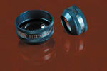
Volk Optical's Digital Wide Field Lens provides the widest field of view available in a non-contact lens. |
|
THE DIGITAL WIDE FIELD LENS
Doctors can now offer both wide field of view and high magnification in one lens with Volk Optical's introduction of the Digital Wide Field lens. The lens provides the widest field of view available in a non-contact lens, paired with the higher magnification required for detailed diagnostic work and imaging.
With a 103°/124° field of view, the Digital Wide field provides visualization similar to a contact lens, without the associated interface solution application and patient discomfort. The .72x magnification produces a more detailed view of the macula and disc, while the design saves time usually spent switching lenses mid-exam.
Volk Optical
www.volk.com
Phone: (800) 345-8655
|

|
|
|
The Epi-K is used to mechanically cleave the epithelium from the Bowman's membrane |
|
EPI-K
Moria has developed the Epi-K, a fully automated system with a disposable head that enables surgeons to perform a new refractive procedure known as epi-Lasik. The Epi-K is an epithelial separator that revolutionizes and extends refractive surgery possibilities.
The Epi-K is used to mechanically cleave the epithelium from the Bowman's membrane leaving a pristine optical zone for laser ablation. Epi-Lasik preserves the structural integrity of the stroma and is expected to minimize discomfort, shorten the length of visual recovery and reduce the incidence of haze associated with other surface ablation procedures, such as PRK and LASEK.
Moria
Phone: (800) 441-1314
www.moria-surgical.com
SEIBEL INTRALASE FLAP LIFTER
Rhein Medical introduces the Seibel IntraLase Flap Lifter. The Lifter is ideal for both IntraLase flaps as well as retreatment with any flap, whether originally created by IntraLase or a mechanical microkeratome. Its unique design maximizes a surgeon's ergonomic mechanical advantage by eliminating the induced torque inherent in conventional flap lifters. It can be operated utilizing a central pivoting technique to further increase mechanical advantage.
The Lifter's dimensions and torsional rigidity are optimized to transmit surgeon force efficiently with minimal artifactual flex. The parabolic shaped tip allows easy initial insertion under flap at hinge. The opposite end of the instrument has a short rounded tip designed specifically to open the gutter and adjacent flap interface at both ends of the flap hinge to further facilitate easy initial insertion of the flap lifter.
Rhein Medical
Phone: (813) 885-5050
www.rheinmedical.com
|

|
|
|
KOWA'S Auto Refkeratometer KW-2000 measures both refractive and keratometry dimensions. |
|
AUTO REFRACTOR/KERATOMETER
Kowa has introduced its Auto Refkeratometer KW-2000, combining a number of features with an intuitive interface. It includes its own printer for instant reports.
The KW-2000 measures both refractive and keratometry dimensions. The keratometry function measures both the center of the cornea and provides measurements in four discrete directions. The unit can accommodate pupils measuring as small as 2.3 mm in diameter and can measure eyes with IOLs. KOWA says the KW-2000 performs its measurements in as fast as 0.07 seconds.
KOWA
Phone: (800) 966-5892
www.medical.kowa.com


