The
Sandwich Technique
New
surgical technique for implanting phakic IOLs for maintenance of deep and
stable anterior chamber.
MANA TEHRANI, M.D. AND PROFESSOR H. BURKHARD DICK
For some time, surgeons have deliberated techniques to better implant phakic IOLs, such as the Verisyse phakic IOL (Advanced Medical Optics [AMO], Santa Ana, Calif.). The Verisyse phakic IOL design (also marketed in Europe as the Artisan IOL) is a stable and predictable refractive correction for nearsighted patients who are not candidates for LASIK. Questions about intraoperative complications have trumped procedural articles raising concerns about the efficacy of phakic IOL implantation.
|
|
|
|
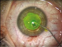
Figure.
A high viscosity OVD, such as HealonGV or Healon5, is injected on the top of the
IOL optic before performing the enclavation. |
|
Intraoperative Management
When implanting the rigid PMMA-model, the surgeon creates a 5.2-mm to 6.2-mm tunnel incision. Currently, all available grasping forceps, including the end-grasping forceps and classical forceps, enlarge this incision. As a consequence, the ophthalmic viscosurgical device (OVD) has a tendency to flow out of the anterior chamber causing chamber shallowing. This outflow can result in endothelial touch by the implant or the instruments used to enclavate the IOL. In turn, this can deteriorate visualization by inducing corneal folds. Moreover, precise enclavation of the phakic IOL becomes increasingly difficult.
To address this complication and facilitate implantation of the phakic IOL, my colleague and I have developed and tested the "sandwich technique." In a study presented at the most recent ASCRS, we achieved a minimization of intraoperative complications such as, flattening of the anterior chamber and endothelial touch. This technique is easy to apply and ensures a safer phakic iris-claw lens implantation by creating and maintaining a deep and stable anterior chamber.
The Sandwich Technique
After performing the primary superior sclerocorneal two-step, self-sealing 5.1-mm to 5.3-mm incision at 12 o'clock, a cohesive medium viscous hyaluronic acid-based OVD is injected into the peripheral anterior chamber opposite the paracenteses. This deepens and allows for a homogenous fill of the anterior chamber. Healon is preferred (AMO) because it is a pure, pH-balanced, iso-osmolar OVD with low endotoxin content.
The phakic IOL is implanted into the anterior chamber and placed into the location where enclavation is intended. After this maneuver, a high viscosity OVD, such as HealonGV or Healon5, is injected on the top of the IOL optic before performing the enclavation (Figure). Thus, the high viscosity OVD is sandwiched on both sides by the medium viscosity OVD. The enclavation forceps grasp the superior haptic and the iris is enclavated via the inferior haptic. To ensure precise enclavation we recommend grasping the iris inferiorly or intentionally moving the lens slightly inferior.
At the end of the surgery, all OVD material is removed by flushing and irrigation with a balanced salt solution on top and in back of the IOL at the end of the procedure. In our technique, the OVD is removed completely with balanced salt solution irrigation. We do not use simultaneous irrigation/aspiration because variations in anterior chamber depth may cause stress on the zonules. Furthermore, the irrigation/aspiration maneuver can also lead to unwanted fluctuations of the iris-lens diaphragma.
We are aware that Healon5 or HelaonGV can be considered more difficult to remove postoperatively, but we recommend these OVDs because of their space-maintaining capability. The high viscosity OVD maintains a safe distance from the implant to the corneal endothelium and positions the IOL closer to the iris. Hence, the enclavation maneuver can be performed more safely.
At the end of the procedure the sclerocorneal incision is checked to be watertight. If a suture is not placed; postoperatively local antibiotic (ofloxacin t.i.d. for 5 days) and steroidal eye drops (prednisolone acetate q.i.d.) are prescribed.
The recovery after implantation is quick with most people returning to their normal activities within the following week.
Additional Tips
We do not recommend placing the high viscosity OVD only at the inner lip of the incision. This maneuver might help to occlude the incision, but can cause an undesirable sudden egress of the OVD as a bolus in some cases.
There have been some concerns about induced astigmatism as a result of a large incision size for phakic IOLs. We do perform a superior sclerocorneal two-step, self-sealing 5.1-mm to 5.3-mm incision when using the rigid model. In a published study,1 we could demonstrate that the induced astigmatism was low: the mean postoperative astigmatism after 6 months was 0.56 D with an axis of 31°. Doubled-angle scatterplot analysis showed a tendency toward more flattening in the vertical meridian.
After applying the �sandwich technique� in more than 150 cases of phakic iris-fixated IOL implantation, no single case of intraoperative anterior chamber flattening or postoperative IOP spike over 25 mm Hg was observed.
Mana Tehrani, M.D. is a resident and research fellow at Johannes Gutenberg-University, Mainz, Germany. Professor H. Burkhard Dick is head physician, professor of ophthalmology for the Department of Ophthalmology, Johannes Gutenberg-University, Mainz, Germany. Neither author has any financial interest in the mentioned products.
REFERENCE
1. Tehrani M, Dick HB, Schwenn O, Blom E, Schmidt AH, Koch HR. Postoperative astigmatism and rotational stability after artisan toric phakic intraocular lens implantation. J Cataract Refract Surg. 2003;29:1761-1766.


