The Reliable,
Yet Resilient, Slit Lamp
This always-important instrument has evolved with the times and continues to do so.
BY ROCHELLE NATALONI, CONTRIBUTING EDITOR
Many instruments were born in response to modern ophthalmic techniques, but few, such as the slit lamp, have preceded them. But rather than rest on its proverbial laurels, the slit lamp, upon which the basic eye exam relies, has evolved with every important trend in modern ophthalmology, from electronic medical records and telemedicine to the need for mobility and physician-friendly ergonomics.
Today's slit lamp does have something in common with its decades-old predecessor: It remains an efficient combination of optics and illumination. What's changed, according to Frank J. Weinstock, M.D., of Canton Ophthalmology Associates, in Canton, Ohio, is the addition of special coatings and filters to improve optics, the introduction of halogen bulbs to improve illumination, the reconfiguration of knobs and switches to improve the ease and therefore speed with which the exam can be performed, and in some cases, the ability to capture and retain or electronically transmit the images viewed.
|
|
|
|
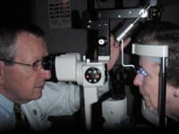
The ubiquitous slit lamp has many new capabilities, including digital image capture and interfacing with other diagnostic instruments. |
|
"We've had photography capability for some time in our practice," said Dr. Weinstock, who is a professor of ophthalmology at Northeastern Ohio Universities College of Medicine. "The most common reason practices like to have this capability is for teaching purposes, but some ophthalmologists use it as a marketing tool by giving patients a picture of their eye. We also use these photos for referral purposes to send a note and picture back to the referring doctor to illustrate the patient's condition," he explained.
Today's medical-legal milieu is yet another reason that slit lamp image-capture capability has caught on, according to Dr. Weinstock. "There's no question that from a medical-legal standpoint if a patient comes in with something that you think in the back of your mind could evolve into a medical-legal situation, it's important to document it," he said.
Joining the All-Digital Revolution
The trends toward digital ophthalmic equipment and electronic medical records have revolutionized the way eyecare practitioners handle patient information. Patient photos, visual field test results, notes and history are increasingly stored in an electronic file, and according to refractive surgeon Daniel S. Durrie, M.D., of Durrie Vision, in Overland Park, Kan., the complete digitization of slit lamp technology isn't that far off. "With all the changes we have had in improving our diagnostic and therapeutic technologies, the one major diagnostic tool that has not essentially changed is the slit lamp. Over the next few years, as we move from the analog to the digital era, I predict we will see changes in the basic slit lamp design. The first step, which has already been introduced, is digital cameras integrated in the slit lamp to allow us to store images in electronic medical records. Future steps will include new image technology such as 3D digital image processing," said Dr. Durrie. "I am sure the basic slit lamp will be with us for years, but there will be a gradual change towards digital equipment just as we have seen in every other field."
Complete digitation of the slit lamp is the ultimate goal, and manufacturers are making strides in that direction. "Instant capture of whatever the practitioner is seeing in the eye is a major advantage for teaching purposes, for legal purposes, for documentation and for referral," said Ricardo Almiron, product manager for medical instruments of a leading slit lamp manufacturer. Almiron pointed out that his company recently introduced new slit lamps that in addition to having more sophisticated optics can be considered totally digital because they have the capability of being easily retrofitted with a digital camera. "You can use the slit lamp as a clinical instrument exactly as the older instruments were, but with the attachment of a small device at the back of the slit lamp, it can inconspicuously be converted to a digital slit lamp with digital capture capabilities of 3.2 megapixels," he said. "Those images can be immediately transferred to a PC, and be downloaded as JPEG or TIFF images and then you can e-mail them, you can print them or you can put them in the patient's report," he said.
Almiron says nowadays a slit lamp with a digital component is a requirement for several reasons starting with documentation. "Documentation is so important today because of the nature of managed care," said Almiron. "You need to be able to seamlessly press a button, get a picture and continue with the exam," he added.
Jacob De La Cruz, vice president of marketing for an ophthalmic instrument company that numbers slit lamps among its products, pointed out that digital capture capabilities are also useful for monitoring changes in progressive diseases such as guttate endothelial dystrophy. "A series of photographs enables physicians to better monitor and measure the progression," he explained.
Real-time image capture and transmission via a video camera is also possible with an appropriately configured slit lamp setup, according to Dr. Weinstock. "The integration of slit lamp findings with specialized computer systems for documentation and following patients is a plus for any practice," said Dr. Weinstock. "The ability to use a slit lamp with a CCTV video camera, on the other hand, is probably only relevant for research and teaching. While the video capability has limited applicability it is nonetheless indispensable for a teacher to have a viewing lens so that students can look in and see what the ophthalmologist sees."
Portability and Equipment Interfacing are Also "In"
De La Cruz said his company has concentrated primarily on improving its optics, but attention to ergonomics has been a priority, as well. "Things like anti-glare coatings and increased light transmission not only improve the quality of the image, they allow for easier, more comfortable prolonged use," he said.
Almiron pointed out that his company has incorporated illumination that is connected in such a way that it simultaneously goes through the slit and through the back (where the digital camera is) producing a very high quality image for printing purposes or for publishing purposes. "We are the trend-setters of a new generation of slit lamps, but I am sure that within one to two years all of the other brands will follow suit," he said.
Handheld slit lamps are another advancement. "These units are useful for patients who have arthritis and can't hold their head up or for situations where equipment mobility is necessary such as in nursing homes or on mission," said Dr. Weinstock. One slit lamp model runs on two AA batteries and can fit into a shirt pocket.
Furthermore, the modern slit lamp's ability to interface with other diagnostic and therapeutic equipment is a benefit for ophthalmologists and patients alike. "The integration of applanation tonometers and fundus lens attachments, the ability to do fundus exams at the slit lamp with noncontact fundus lenses, and the capability of combining the slit lamp technology with Nd:YAG laser technology for trabeculoplasty have been wonderful advances," said Dr. Weinstock. The ultimate luxury though, he says, is having the same model of slit lamp throughout a practice. He suggests that while it may seem impractical to upgrade all slit lamps simultaneously, "having all of the knobs and buttons in the same place, makes things so much quicker and easier."
Worth Every Penny
A bare-bones slit lamp can be purchased for as low as $3,000, while a state-of-the art setup with the best optics and the capability to interface with all of the available imaging bells and whistles can cost as much as $12,000. "But not every practice needs a top-of-the line unit," says De La Cruz, who puts the average ophthalmologist's cost for a single slit lamp at $5,000 to $7,000.


