Posterior Corneal Transplants
The descemet stripping automated endothelial
keratoplasty (DSAEK) procedure offers a new alternative for endothelial disease
sufferers.
MARK S. GOROVOY, M.D.
For endothelial disease sufferers, penetrating keratoplasty (PK) results can be uncertain. While the anatomical results (i.e., clear cornea) are excellent, the visual results judged by speed, quality and predictability are poor. Globe integrity is at risk with a long-term 5% ruptured wound rate, which can often result in total vision loss. In my practice, for previously failed PKs and endothelial disease sufferers — Fuchs and pseudophakic bullous keratopathy (PABK) — PK is no longer the procedure of choice.
In order to make corneal transplant results for these types of patients safer and more predictable, a new surgical technique, DSAEK, has been developed. My DSAEK procedure has evolved from the revolutionary work of Gerrit Melles, M.D. Additionally, Mark Terry, M.D., and Francis Price, M.D., have been instrumental in the procedure's evolution. The overall goal of DSAEK is to eliminate full-thickness trephination and replace only the diseased Descemet/endothelial complex. DSAEK eliminates all manual dissections, affording the patient an opportunity for a rapid visual recovery, which can result in a typical 20/40 best spectacle-corrected vision by 4 to 6 weeks postoperatively.
|
|
|
|
Figure
1. The Moria ALTK artificial chamber cut with 300 μm head removes donor tissue. |
DSAEK Procedure
When removing the donor's cornea, I utilize the Moria automated lamellar therapeutic keratoplasty (ALTK) system (Antony, France) with a 300-μm CBm head (Figure 1). The remaining posterior cornea is cut endothelial side up with a 9-mm punch. This large donor size increases the number of donor endothelial cells and eases the surgical centration of the donor tissue up against the recipient cornea.
On the recipient's eye, I create several diamond paracenteses to help with anterior chamber access. A 9-mm epithelial trephination mark outlines the descemet scoring. Descemet's membrane is scored and peripherally stripped for at least 180Þ with my irrigating stripper. Through a 3.2-mm temporal clear corneal keratome incision, an irrigation and aspiration (I&A) handpiece is introduced into the anterior chamber and the loose-scored Descemet's membrane is aspirated and removed from the 9-mm central cornea. The corneal incision is then enlarged to 5-mm.
After applying a small amount of viscoelastic on the donor endothelium, I overfold it (60/40) and insert the endothelial lamella into the anterior chamber with curved tying forceps. Two 10-0 nylon sutures close the corneal wound and facilitate a deep anterior chamber with irrigation. I observe the folded donor tissue to ensure it unfolds with the stromal side up.
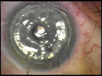
Occasionally, this unfolding process can be aided with a Sinskey hook or even an I&A. The unfolding requires a deep anterior chamber, and I avoid the endothelium. The tissue is then centered by using finger ballottement at the limbus easily pushing the donor tissue in place. The anterior chamber needs to be only partially deep for this movement. Lastly, a large air bubble is injected under the donor tissue, filling close to 100% of the anterior chamber (Figure 2).
Upon finishing the procedure, dilating drops are applied, the eye is patched, and the patient remains supine in the postoperative holding room for 30 to 60 minutes. A slit lamp exam verifies acceptable donor positioning and enough air is burped out of the paracentesis to prevent pupillary block. Before the patient is discharged, a follow-up appointment for the next morning is scheduled.
|
|
|
|
Figure 2. An air bubble under the donor tissue
is created filling the anterior chamber of the recipient's eye. |
|
Postop Care
Ideally, on the first day postop, the donor corneal button is well-centered and relatively clear. Corneal edema usually clears by the seventh day postoperatively if there is good donor/recipient adhesion. I find this occurs in approximately 75% of patients.
The remaining 25% of patients will have gross corneal edema with a discernible donor gap noted on slit lamp exam. If the donor tissue is decentered, I look at the patient's eye under a microscope, I re-center the tissue and refill the anterior chamber with a large air bubble. They remain supine for 30 minutes and the air is partially burped out of the anterior chamber to again prevent pupillary block.
A second postop air bubble is
rarely required. If on day 1 the cornea is grossly edematous but the tissue is well-centered
with no gap or a minimal gap, I often follow the patient weekly and will insert
a new air bubble if I feel the gap and edema does not improve. I have observed two
patients who spontaneously cleared up at postop week 7 with excellent donor endothelial
cell counts — greater than 2000 — using a specular microscope. I inform
my patients with normal retinal potential that over 90% of them should recover at
least 20/40 best spectacle-corrected vision
postoperatively by 4 to 6 weeks.
Slit lamp exam of these transplants show clear, smooth interfaces with corneal thickness
ranging from 575 μm to 800 μm. Excellent vision is unrelated to final
pachymetry.
Patient Types
I presently perform the DSAEK surgery as a stand alone procedure. Patients with Fuchs undergo lens surgery with a post-cataract IOL 4 to 6 weeks prior to DSAEK. PABK patients with anterior chamber IOLs have them replaced with scleral-sutured pseudophakic IOLs 6 weeks prior to DSAEK.
I never recommend elective surgery on the second eye until the first is the better eye. This can often take longer than one year for PK eyes, but only 2 to 3 months in my DSAEK patients — even those with 20/40 preoperatively. Rigid contact lenses, surface disease, wound rupture, relaxing incisions, compression sutures, broken sutures, suture abscesses and irregular astigmatism, are all adverse advents of the past when utilizing the DSAEK procedure. Of course, I am still performing corneal transplants, so rejection and steroid-induced glaucoma are not eliminated. Additionally, cell counts may invariably decline over time, but the DSAEK can be repeated to address this issue.
DSAEK surgery has completely changed the way I approach corneal transplants. Patients no longer have to suffer from the long, unpredictable and often poor visual recovery of PK. Because of the confidence I have in DSAEK, I can confidently recommend surgery sooner to many of my patients.
Mark S. Gorovoy, M.D., practices at his Gorovoy MD Eye Specialists' office in Ft. Myers, Fla., and teaches a monthly DSAEK course. He can be reached at (239) 939-1444 or Bbarr@gorovoy.com.


