spotlight on technology &
technique
At Up to 25 Frames/Second, this Ultrasound is Ultrafast
The capability of OTI's HF35-50 to record dynamic images increases your diagnostic precision.
By Kay T. Adams, Managing Editor
Ultrasound imaging technology has dramatically changed the way we can see inside the human eye without invasive procedures. Now, with the HF35-50 High Frequency Ultrasound system from Ophthalmic Technologies Inc., you can perform high-frequency ultrasound biomicroscopy of the anterior segment and high-resolution ultrasound of the posterior segment.
The HF35-50 has an 18.5-mm-wide scanning field that shows the entire anterior segment, limbus to limbus, affording a cross-sectional view of the cornea, anterior chamber and both leaves of the iris. The system captures static images as well as real-time, dynamic recordings that show movement within the anterior chamber.
You can view the dynamic recordings start to finish, like a movie, or view images frame by frame. By using the fast-forward and reverse functions, you can analyze each frame to more clearly discern pathologies or study structural relationships.
All images are stored on the hard drive, so they can be accessed at any time, burned onto a CD or DVD, or viewed through a local network. If you want to use images for a presentation, for example, all you have to do is export them in TIFF or PDF format.
Using the System
Performed by you or a skilled technician, the whole imaging procedure takes approximately 5 minutes. The system comes with three immersion cups to match eye openings. A foot pedal controls the image gain and size of the dynamic imaging "movie."
From a practice management point of view, the dynamic recording capability is a benefit because it lets you maintain your patient flow and the amount of time you spend with patients. Performing a standard anterior segment examination frame by frame can be time-consuming, but with the HF35-50 you or a technician can quickly capture the images, store them in the system's database, and view them at a later time to study features and pathology more closely.
Patients may also gain a clearer understanding of their diagnoses by seeing the dynamic imaging movie on-screen.
Combining Components
The A-scan and B-scan capabilities are available on PC and Macintosh platforms. With both you have software compatibility, which means you can mix and match components, for example, 35/50 MHz anterior segment, 12/20 MHz posterior segment B scan, 12 MHz 3D posterior segment B scan, and 13 MHz A scan. All scans are saved in a common database on a single computer. Upgrading is straightforward; you can start with a few components and add others later.
The HF35-50's capabilitiy for imaging the entire anterior segment, from the cornea all the way to the anterior vitreous, is especially valuable for detecting anterior-segment tumors or complications resulting from a vitrectomy.
The instrument also has a wide scan angle, 38 degrees, and a deep scan depth that can reach 15 mm from the transducer to view angle to angle and sulcus to sulcus.

|

|

|
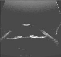
| Image taken before iridotomy shows the iris bows backward. | Image captured 20 minutes after iridotomy shows the flat iris. | Image captured 20 minutes after iridotomy shows the iridotomy site. |
Feedback from the Field
Carol L. Shields, M.D., who practices at Wills Eye Hospital in Philadelphia, Pa., says that the HF35-50's panoramic view of the anterior segment can't be obtained with any other anterior segment ultrasound system. Also, being able to view the entire patient examination in a continuous format lets her note interesting features in relationship to other structures.
"This is especially important, for example, when evaluating the relationship of an iris cyst to a solid iris mass that might be a little bit eccentric. The relationship of iris dialysis can be more clearly studied in its extent as the periphery of the iris is scanned," she says.
The system's ability to image the full anterior segment, ciliary body as well as the peripheral retina can be critical in determining therapy, Dr. Shields points out.
"We recently had a patient sent to us from another state with a diagnosis of an iris nevus. Using the OTI system, we were able to image the pigmented iris lesion, but we also noted a large nodule behind the iris of 3.5 mm in thickness, which is consistent with possible early melanoma, and this was not visible on clinical examination. Using the digital movie capability, we were able to visualize the extent of the iris component as it related to the ciliary body component," Dr. Shields explains.
Improvements to the system may be on the way, she says. "OTI is working on improving image resolution, which would allow delineation of the iris pigment epithelium from the iris stroma." Another limitation is related to the physics of how sound bounces off the cornea. "In some cross sectional cuts, the mid-cornea loses its echogenicity whereas the central cornea, peripheral cornea, and limbus maintain their echogenicity," she explains.
Richard Rosen, M.D., who practices at The New York Eye and Ear Infirmary, also uses the HF35-50 system. "It's another step up in terms of what we need to do as opthlamologists," he says.
"With conventional ultrasound, you take a single slice through the eye, but you may miss the relevant pathology or do it at an angle that doesn't make sense. By capuring the examination dynamically, you can answer many questions about the anterior pathology very quickly. You can see into the recesses of the angle or measure the true width of the eye, which is important in intraocular lens placement."
To make the most of the technology, Dr. Rosen prefers to perfrom the ultrasound testing himself, rather than delegating the task to a technician.
"In ultrasound the issue is determining what's artifact and what's signal," he explains. "So by doing the examination myself, I can understand exactly what is happening. There is a certain learning curve, but not like with vitrectomy. It's important for physicians to learn the skill so they can understand what can be done with the instrument."
Additional Features
The HF35-50 system also offers these features:
- compact, lightweight interchangeable 20 MHz, 35 MHz or 50 MHz probes
- measurements of angle to angle, sulcus to sulcus, angle in degrees, corneal thickness, anterior chamber depth, lens thickness and scleral thickness (Many of these measurements are crucial to outcomes if you're implanting the new phakic IOL or other refractive implants.)
- post-processing adjustment of gain, TGC, and contrast
- compatible with inkjet and laser printers.
- You can also combine the HF35-50 with the OTI-Scan AB + 3D system. The resulting combination instrument would contain the high-frequency probe, B scan probe (with integated 3D scanning), and diagnostic A probe, giving you the ability to diagnose both anterior and posterior segment conditions.
For More Information
To find out more about the OTI HF35-50, call (888) 517-4444, e-mail info@oti-canada.com, or visit www.oti-canada.com.


