Expanding Applications for Ultra Widefield Technology
Optos is extending the utility of
the Optomap Panoramic200 to include fluorescein angiography.* Combining these diagnostic methods can show more than either could alone.

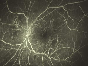
Small white spots represent retinal hemorrhages in diabetic retinopathy.

Ultra widefield view shows the retinal periphery, making it possible to see pathology not imaged by fundus cameras.

Despite extensive panretinal photocoagulation for diabetic retinopathy, areas of fluorescence indicate continued neovascularization in this patient.

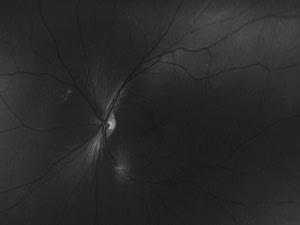
This Optomap images shows a normal fluorescein angiography.

In this image, small chorioretinal infiltrates indicate posterior uveitis.
*Pending FDA approval.
Q&A
Question: Would you recommend the Optomap exam as a substitute for the dilated fundus exam?
Answer: Physicians must determine for themselves if dilation is necessary. Ideally, every patient would have a thorough dilated manual indirect exam and an Optomap, enhancing disease detection and providing permanent documentation.

Question: Do you store Optomap images on a computer or do you print a hard copy for each patient?
Answer: Sometimes I'll print images for patients, but hard copies don't have the same diagnostic value as digital images. The Panoramic200 comes with CD-burning software and a hard drive that can store each image in the patient's file. You can compare new images with previous examinations instantly or transfer digital images to your colleagues. Versatile image-sharing and recall are among the most appealing features of the Optomap system.

Question: Does the Optomap Panoramic200 require special modifications to work with fluorescein angiography?*
Answer: The only necessary modification is installing a blue laser. The standard Optomap system uses red and green lasers to provide information about different retinal layers, including the presence of disease or pathology. Whereas the green laser scans superficial retinal layers, the red laser shows structures deep to the pigment epithelium, including choroidal arteries and veins, or as illustrated by the images to the right, abnormal structures (see "Separating Retinal Layers"). Blue lasers pick up yellow-green light emanating from intravenous fluorescein in the retinal vessels, providing a detailed image of the retinal blood supply and highlighting any areas of fluorescein leakage (see "Pinpointing Pathology").

*Pending FDA approval.
| Separating Retinal Layers
A. |
B.
|
C.
|
| The first Optomap image (a) shows a choroidal retinal nevus superornasal to the optic disc. Green (b) and red separation (c) images confirm that the nevus is confined between the pigment epithelium and choroidal retinal layers. |
|
Pinpointing Pathology
Optomap fluorescein angiogram showing extensive leakage near the optic disc. |


