spotlight
on technology & technique
Retinal Anatomy in 3-D
Fast scans and sophisticated software now make it easy to examine macular tissue from the inside out.
By Christopher Kent, Senior Associate EditorHow the RTA Works
As technology has evolved, it's become possible to map and display an increasing amount of anatomical detail inside the eye. Now a more advanced use of the data collected by Talia's RTA (Retinal Thickness Analyzer) is making it possible to see retinal anatomy in entirely new ways, with a remarkable level of interactive control.
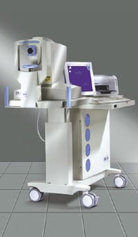
The RTA is an imaging and diagnostic tool used for screening, diagnosis and follow-up of retinal pathologies, including macular holes and cysts, diabetic/macular edema, AMD and glaucoma.
A scanning laser ophthalmoscope maps the topography of the optic disc and thickness of the posterior pole, fovea and/or peripapillary region. This data is overlaid on a fundus image to clarify location and allow for deviation analysis in the future. (Also, see "How the RTA Works".)
Going Interactive
In the Spring of 2003, with the release of RTA version 4.20, Talia launched its "modular concept," allowing customers to purchase optional modules that enhance the capabilities of the RTA. The first module made it possible to store the fundus images taken by the internal camera. Talia premiered a second module later in the year: The 3-D Anatomy Imager.
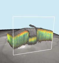
The RTA has always been able to produce a 3-dimensional surface map representing the scanned retinal/disc data. However, the 3-D Anatomy Imager goes much farther, assembling the data from the series of 16 sequential cross-sections of retinal thickness into a 3-D "volume visualization" -- a moveable, "sliceable" block showing all the details of the 3 x 3 mm scanned area in three dimensions. Also referred to as a "voxel" (a 3-D version of a pixel) reconstruction, this representation contains far more detail than the earlier 3-D simulation.
The 3-D block appears "suspended in the air," allowing you to manipulate the image in several ways. You can:
- rotate the 3-D section 360° in any direction
- zoom in or out
- display the data in black and white, or color-coded using an algorithm to enhance the inner structure of the retinal layers
- adjust appearance using variable delineation to emphasize different details
- measure distances and angles of your choice
- slice the 3-D section anywhere along the x, y or z axes to see conditions inside the tissue. (The axes rotate with the section.) You can see the view from either side of the slice -- i.e., from the front or back.

|

|

|

|
|
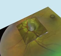
The RTA's 3-D Anatomy Imager module lets you rotate and slice 3-D representations of the scanned area of the retina, along the x, y or z axes. Left to right: Diabetic macular edema; glaucoma; a macular hole (sliced to reveal internal structure); optic nerve head. |
|||
Feedback From the Field
Pamela A. Weber, M.D., is a retinal specialist practicing on Long Island in New York. She's worked with the 3-D module for several months. "The RTA is a really great product, with extensive clinical application," she says, "but we're just learning how to apply the new 3-D Anatomy Imager. It looks very promising for imaging macular pathology, particularly diabetic retinopathy, and maybe even subretinal neovascularization associated with macular degeneration. It's very good for imaging macular holes."
Asked how good a diagnostic tool the 3-D Imager will be, she says it's hard to know. "We're not used to looking at three-dimensional pictures moving in space. Sometimes I see interesting things, but I'm not sure what I'm looking at. We need to scan numerous patients with known pathology, so we'll recognize the diseases in 3-D."
Richard Rosen, M.D., a retinal specialist at the New York Eye and Ear Infirmary in Manhattan, says the new module gives him a much clearer picture of what's going on in the retina. "The original instrument showed us that the retina was thickened, but you couldn't tell if there was a cyst in the middle of that area. The new 3-D Imager reveals those details.
"Once you've taken the picture, you can adjust the image to clarify anomalies. You can slice it along any of six axes, so you can look at the same lesion from six perspectives. You can also import images from other modalities, such as fluorescein photographs, so you can see leakage in the fluorescein image and use the 3-D Imager to visualize the physical components. (The RTA automatically aligns the images.)
"The 3-D Imager is almost in the domain of the vitreoretinal surgeon because a lot of what you see is potentially surgical," he notes, adding that the RTA and new module are very user-friendly. "The technician doesn't have to be sophisticated to get good, consistent information."
Dr. Rosen says that one advantage of the RTA is the large map of the retina it produces by stitching together multiple scanned segments. "Currently, the 3-D Imager only shows one area at a time, but Talia is developing the software to stitch together the 3-D components.
"One of the strong points of the RTA has always been the extensive package of features within the instrument -- it can analyze retina, diabetics and glaucoma. In an office setting it offers a lot of value, and this module adds to the substantial capabilities of the instrument."
Only the Beginning
The RTA, like most imaging tools, is sensitive to media opacifications such as cataracts. Nevertheless, the RTA has established a strong track record, and revealing new levels of information can only have a positive impact on diagnostic capability. Of course, being able to show a 3-D scan to patients is also likely to have a significant impact, in terms of helping them to understand their condition, encouraging compliance -- and impressing them with your high-tech practice.
To learn more about the RTA, contact Talia Technology at (800) 214-2030, or visit the Web site at www.talia.com.
|
How the RTA Works |
The RTA can translate the resulting information into a number of different color-coded maps showing either retinal thickness or disc topography; it can also produce a color-coded map showing deviation from a normative thickness database. (A standard RTA scan of the central macula can detect thinning around the fovea indicative of early glaucomatous damage -- often before changes in the optic nerve head become evident.) |


