Using Wavefront Aberrometry As a Primary Diagnostic Tool
Wavefront technology has
enormous potential to become the next standard for routine refractive testing and diagnosis.
By Daniel S. Durrie, M.D., Overland Park, Kan.
|
|
|
|
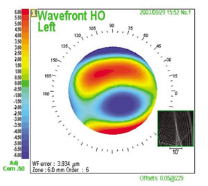
Wavefront map showing coma. |
|
|
|
|
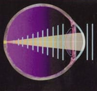
Representation of an ideal wavefront.

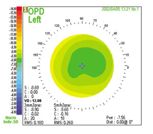
Optical Path Difference map showing keratoconus.

Wavefront map showing trefoil in patient with poor night vision.

Internal Optical Path Difference map showing a torqued IOL.
Our increased understanding of higher-order aberrations has helped us develop newer, better refractive surgery techniques. And yet, we still test our patients' visual acuity with phoroptors. What's wrong with this picture?
Wavefront technology isn't just for patients who want laser vision correction; this technology offers all patients a more comprehensive vision analysis than a traditional phorpotor examination. I do wavefront analysis for every patient at every visit because it's simply a better diagnostic tool.
How Wavefront Aberrometry Works
In an ideal optical system, light rays would enter the eye, focus on the retina and reflect a perfect flat waveform. But none of us is perfect. With a wavefront aberrometer, we can see how defects in the lens or the cornea distort light rays as they enter the eye, forming some other non-linear pattern. Wavefront technology lets us examine lower-order (sphere and cylinder) and higher-order optical aberrations and understand how they affect visual acuity.
Currently available wavefront aberrometers use several types of sensors to capture and compile data. Tscherning wavefront technology projects a grid on the retina; Hartmann-Shack bounces a light wave off the retina and uses a lenslet ray to read the wave as it exits the eye. Tracey technology projects single points of light on the retina sequentially to measure wavefront error. The Marco 3-D Wave uses dynamic skiascopy to refract the entire pupil and produce a map of the patient's eye, showing the relationship between a patient's optical aberrations and his visual symptoms.
Diagnostics for Difficult Cases
The Marco 3-D Wave combines wavefront technology with three other diagnostic technologies to evaluate a patient's total visual system. I use this device to help patients who have postsurgical problems, as well as those with long-term, previously untreatable refractive conditions.
Post-LASIK effects. Since I started using wavefront technology I've become so familiar with the symptoms of coma and spherical aberration that I can usually predict what a patient's wavefront analysis will show by his symptoms alone. And even though many of these patients aren't interested in additional treatment, they want me to identify why they're having problems. When these patients come to me for diagnostic wavefront aberrometry, I can tell them, "This is why you see how you do."
Keratoconus. Wavefront technology is well-suited for diagnosing and following refractive changes caused by corneal pathologies such as keratoconus. Different pathologies have distinctive wavefront maps that we can learn to recognize. Eventually, devices will be able to use wavefront data to identify pathologies that may otherwise be missed.
Night vision problems. Unlike traditional topographers, the Marco 3-D Wave takes refractions at multiple pupil diameters. After measuring a 3-, 5- and 7-mm pupil, you may discover a patient is 2 diopters more myopic at the periphery of his dilated pupil. Sometimes I'll look at a patient's results and ask him if he has trouble seeing at night. If he says, "Gee, I have all my life," I can use the zonal refractive feature of the Marco 3-D Wave to explain why he has poor night vision, and take the opportunity to discuss treatment options.
Intraocular lens (IOL) complaints. Previously, when patients returned after cataract surgery complaining of continuing trouble with their vision, it was often difficult to isolate the exact cause of their problem. Now we can use wavefront technology to subtract the refractive status of the cornea from a patient's total measured visual defects to calculate what visual defects are coming from the IOL. In the future, we may be able to use this same process to determine whether cataracts are visually significant.
Wavefront Technology's Future Role
Diagnostically, we're just scratching the surface of wavefront technology's potential benefits. I wouldn't be surprised if wavefront aberrometry eventually replaces the phoroptor as the standard for diagnostic refraction. Wavefront technology provides previously unavailable information about our patients' visual systems, enabling us to diagnose and treat hitherto poorly understood problems. Moving to a wavefront-driven practice will open exciting new doors and help our patients see better than ever before.
|
Dilation Gives the Whole Picture |
||||
A recurring question among wavefront users is, "Do I need to dilate the pupil for an accurate wavefront reading?" The answer is definitely yes, especially if you want to find out how laser eye treatment will affect a patient's night vision. Because refractive surgery changes a patient's vision forever, you should always use the best available data when planning your procedure. And the best data come from a dilated pupil. I use dilated wavefront information for excimer laser treatment because it provides more information about optical aberrations, including those occurring in the periphery of the pupil. Even if I'm working within a 6-, 6.5- or 7-mm treatment zone, I still collect as much peripheral data as possible. More peripheral data translates into better night vision for my patients. |
|
Linking Wavefront Aberrometry And Lasers |
The newest development in wavefront technology is wavefront-guided excimer laser correction. At first glance, wavefront-guided LASIK is deceptively simple: You just export the data from the aberrometer to a laser and let the laser use wavefront data to reshape the cornea. However, because laser ablation changes a patient's vision forever, you must take the time to assess the aberrometer's raw data. Many people go straight to the color wavefront map without looking at the lens array information. I like to look at the raw data, different pupil diameters, the ratio between lower- and higher-order aberrations, the total root mean square and the 3-D map. These data help me understand how laser ablation will change the shape of my patient's cornea. Another critical element for bridging aberrometry and the excimer laser is the human element: You. You can delegate diagnostic wavefront analysis to technicians, but not wavefront analysis intended to guide a laser. In this situation, measurement is just as important as the procedure itself, and it requires your expertise. With all of these checks in place, your practice is ready to link aberrometry and the excimer laser -- and offer patients truly customized vision correction. |


