Uncovering Visual Complexity
Precision wavefront technology joins other state-of-the-art diagnostic instruments in a single integrated
system. Here's how it can make a
difference in your practice.
By John F. Doane, M.D, Kansas City, Mo.
We've made great strides in refraction and vision correction, but only recently, with wavefront technology, have we truly uncovered the complexity of the human visual system. Automated instruments make it easier than ever to apply our newfound knowledge in a clinical setting.
The Marco 3-D Wave integrates refractive analysis, corneal topography, Optical Path Difference (OPD) and wavefront analysis in one device that provides:
► Accurate, detailed and accessible data
► Easy-to-use integrated tools that trim testing time
► Tangible clinical advantages over similar (separate) xxxx devices.
And because the 3-D Wave offers wavefront technology, we can analyze a patient's complete visual system, including higher-order aberrations.
|
|
|
|
|
|
New Perspectives on Visual Complexity
Visual acuity is the sum of a complex interaction among light rays and the cornea, the crystalline lens and the retina. The slightest irregularity in any optical structure disrupts the whole visual system, causing refractive errors and visual distortions.
With the help of phoroptors, and other traditional tools, we routinely diagnose refractive errors that cause lower-order aberrations such as myopia, hyperopia and astigmatism. We easily remedy these spherocylindrical errors by refocusing the light entering the eye with eyeglasses or contact lenses or by physically changing corneal curvature with refractive surgery.
However for a minority of patients, 10% or more of their vision problems are caused by irregular astigmatism, which can't be improved with eyeglasses or contact lenses. We didn't have the technology to correct or even measure the optical aberrations that cause irregular astigmatism -- until wavefront technology.
Wavefront technology measures the complete refractive status of an optical system by comparing the configuration of light rays reflected from the eye with an idealized, flat wavefront perpendicular to the eye's surface. Special sensors translate the optical path distance or wavefront error into identifiable components called Zernike polynomials. Optical errors that were once grouped together as irregular astigmatism are now expressed as higher-order aberrations named according to degree of angular frequency and radial order.
|
|
|
|
Corneal topographic map showing traditional with-the-rule astigmatism. |
But what does all this mean for the average patient? Put simply, wavefront technology lets us define refractive error on an individual basis. For example, for a 10-diopter myope, an average 97% to 99% of the light-bending problem affecting his vision is sphere and cylinder. One percent to 3% is higher-order aberration. For a lower ametrope, 70% to 90% of the problem is sphere and cylinder, and 10% to 30% higher order aberration. Using wavefront aberrometry to analyze vision lets us personalize the outcomes, not just correct all higher-order aberrations.
Increasing Practice Efficiency
The Marco 3-D Wave has been a valuable addition to our practice. I call it the "diagnostic dynamo." One push of the button takes near-simultaneous keratometry, topography, refraction and wavefront measurements. You can get a report that includes axial, elevation and OPD maps, higher-order breakdown and numerical data for each eye. The system lets you explore different screens and pull as much or as little information as you want to print on a single printout.
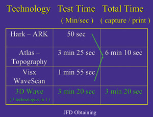
An integrated instrument is faster, as well. As illustrated by the chart on page 6, it takes 50 seconds from pushing the button on my autorefractor to having a hard copy in my hand. The topographer takes 3 minutes and 25 seconds, and the WaveScan (VISX) takes 1 minute and 55 seconds. The total -- 6 minutes and 10 seconds -- is nearly twice the 3 minutes and 20 seconds it takes to get all the same data with the Marco 3-D Wave. And the 3-D Wave offers nine measurements to help us plan our patients' treatments.
Our technicians love Marco 3-D Wave so much that if I have them use another device they ask to go back to the Marco. Their enthusiasm for the device encourages good understanding and use of the technology, which means better testing for patients.
|
|
|
|
|
|
Clinical Advantages
In my practice, our experiences with the 3-D Wave have been outstanding. The 3-D Wave's autorefractor is the best we've ever worked with in our office. It has the best mean and the best and tightest standard deviation compared to my gold standard of maximal pushing-plus manifest refraction.
One thing I appreciate about the 3-D Wave compared to other autorefractors and wavefront analyzers is that this device has the best non-accommodative target on the market. With other analyzers, you're looking at the patient's pupil and seeing him accommodate. With the Marco 3-D Wave, the patient has a much more stable pupillary response -- accommodated the whole time or not accommodated. Data gathered by the 3-D Wave is virtually identical to data we get using maximal pushing-plus manifest refraction.
From a data standpoint, we've had excellent results for our patients. To compare the Marco 3-D Wave to subjective refraction, we did a study in our practice. In 50 eyes measured using the 3-D Wave, the sphere's mean was -4.20D, compared to -4.17D for subjective refraction. The cylinder's mean was +0.92, compared to +1.02, and the axis was 084, compared to 075. The standard deviations were very close in the two methods.
Detailed Accuracy With One Button
By giving us four critical data points, including wavefront analysis, in about 3 minutes, the Marco 3-D Wave changes the way we collect and view patient data. In my practice, the device saves time, improves outcomes and helps us make better and faster treatment decisions.
|
Understanding Higher-order Aberrations |
Optical aberrations are deviations from stigmatic, or perfect images that occur when light rays pass through defects in the cornea and the lens. In ophthalmology, we typically talk about field curvature, distortion, spherical aberration, diffraction, chromatic aberration and coma. Previously, these aberrations were difficult to understand or visualize. However, we can now use wavefront technology to detect higher-order aberrations, and perhaps more importantly, to choose treatment that prevents us from introducing additional optical defects. One common higher-order aberration caused by traditional LASIK is spherical aberration. As light rays pass through a convex lens, centrally located or paraxial rays focus directly on the fovea. Rays passing through the lens periphery become distorted, eventually converging just anterior to the retina and producing a less-than-ideal blur circle. Under normal circumstances, corneal shape -- flatter on the periphery and curved in the middle -- compensates for spherical aberration by redirecting light toward the fovea, producing a clear, sharp image. Sometimes changing a patient's corneal shape with surgical ablation inadvertently increases spherical aberration. This may be good news for a 40-year-old patient, who may gain greater depth of focus, but the same change in a 20-year-old patient can lead to night myopia. |


