spotlight
on technology & technique
Data in Stereo
Combining two sources of information can reveal more than either one alone.
By Christopher Kent, Senior Associate Editor
As technology advances, it's become possible to visualize and measure ever tinier structural details inside the eye. Sometimes, however, the most helpful information doesn't come from uncovering new information, but from finding new ways to combine and cross-analyze existing sources of data.
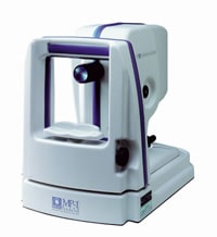
Now, a new instrument from Nidek makes it possible to precisely overlay visual field information obtained by perimetry on a non-mydriatic photograph of the retina. Combining these two data sources lets you see where visual field defects coincide with visible structural anomalies.
|
|
|
|
The MP-1 from Nidek
accurately overlays data from perimetry and fundus photography. |
|
Multi-Level Data
The MP-1 Micro-Perimeter from Nidek performs both digital perimetry and non-mydriatic fundus imaging; high-speed tracking software makes it possible to accurately overlay the two resulting maps. In addition, the MP-1 can perform special functions such as delineating scotomas. These abilities make the instrument exceptionally useful for mapping visual function before, during and after treatment.
Visual field. The MP-1 performs microperimetry using selectable target strategies and user-programmable parameters that allow you to choose target number, intensity, shape and size, as well as position strategies and foreground/ background colors. You can also perform fully automated exams using selected strategies based on standard Goldmann targets, operator-defined targets or bitmaps.
Retinal imaging. The MP-1 uses infrared light to generate real-time, non-mydriatic 45° images of the retina, displayed on a monitor during the exam. (The use of infrared light ensures patient comfort and cooperation.) The instrument captures a full color digital image at the beginning or end of the exam.
Tracking eye movement. High-speed tracking software monitors fundus movement while the patient is focusing on the fixation target to ensure that the anatomic landmarks revealed in the fundus photo are precisely aligned with the sensitivity maps generated by the perimeter. (The tracking system can also produce a graph showing the patient's eye movement during the exam, along with a quantitative assessment and analysis of fixation.)
Delineating scotomas. The MP-1 can accurately detect and delimit "blind" areas on the retina. The instrument projects a light onto the retina at a test location; the light then moves radially away from that location until it becomes visible to the patient. The resulting map can also be laid over the fundus photograph.
A Host of Uses
Data collected by the MP-1 can help you diagnose AMD, CNV, diabetic retinopathy, maculopathies, scotomas and similar problems, as well as evaluate the impact of retinal therapies such as PDT, TTT or DEP. According to Nidek, the reliability of the MP-1 system makes results highly reproducible. And, as you know, both fundus photography and perimetry are reimbursable.
For more information about the MP-1, contact Nidek Technologies America at (888) 382-5064 or info@nidektech.com, or visit www.nidektech.com.


