Creating a "Virtual Biopsy"
Improvements in technology make it possible to "see" more internal tissue detail than ever.
BY CHRISTOPHER KENT,
SENIOR ASSOCIATE EDITOR
In the movie Fantastic Voyage, scientists shrink themselves to microscopic size and travel through the human bloodstream to examine the condition of a patient's body. We'll have to wait a few more years to use that particular technology, but in the meantime, advances in computers and instrument design are making it possible to approach that level of tissue observation -- at least in the eye -- without having to surgically invade the tissue.
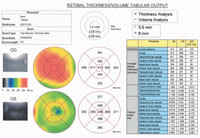
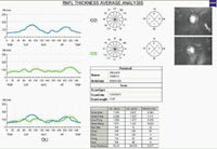
The newest version of the OCT instrument from Carl Zeiss Ophthalmic Systems is a case in point. The OCT3 scans tissue using optical coherence tomography -- essentially the same technology as ultrasound, but using light instead of sound. Measuring with light allows the OCT 3 to create far more detailed images than would be possible with sound. In fact, this is the only technology that can measure and display individual layers in macula, RNFL and optic nerve head tissue, providing a complete cross-sectional view of retinal structure.
What's most remarkable about the OCT3 is that it far surpasses the detail captured by earlier models and competing technologies. In fact, the resolution achieved by the OCT3 is so impressive that surgeons are referring to the images as "virtual biopsies" and "real-time histology."
A big step forward
The OCT3's advantages over previous models include:
|
|
|
|
Above: The new OCT3 from Carl Zeiss Ophthalmic Systems. |
|
|
|
|
|
A scan made by the OCT2 (top) compared with a scan from the OCT3 (bottom) with 10 times the resolution. |
distinguish seven or eight layers. (See images, right.) The OCT3 is the only instrument to image the ganglion layer in addition to the RNFL. In fact, the OCT3 shows details never seen before except in histology, such as the umbo.
This is possible because the OCT3 measures 10 times as many data points (achieving 10 times the resolution) of the OCT1 or 2. The OCT3's resolution is 100 times greater than standard ultrasound.
You don't need to dilate. The OCT3 can scan through a pupil that's only 3 mm wide. (Previous versions of the OCT, and most competing technologies, require at least 6 mm.)
Reimbursement is now available for retinal use. Use of the OCT3 for retinal analysis is now reimbursable in 20 states. This means that the multiple capabilities of the OCT3 can have a much greater impact on your practice's bottom line.
Other features
The OCT3 offers several other advantages, compared with its predecessors and competitors:
- The OCT3 acquires scans 5 times faster than earlier models, making "simultaneous" combination scans possible. It can take six circle scans at once, eliminating registration issues between separate scans.
- The internal computer is faster and has more memory; it can store 10 times the data of previous models.
- The instrument has a smaller profile; you can operate it without a special table. (The table provided with the instrument is half the size of the table needed for the original OCT, and 3/4 the size of the table that came with the OCT2. It's also wheelchair accessible.)
- Basic controls are located on the scan head.
Comments from the field
Dr. Philip Rosenfeld, M.D., Ph.D., of the Bascom Palmer Eye Institute at the University of Miami School of Medicine, has been using the OCT3 to examine retinal patients.
"The OCT3 is an enormous step forward in our ability to evaluate the retina and vitreoretinal interface in situ," he says. "Never before have we had the ability to see the retina with this kind of resolution. The OCT3 should further our understanding of retinal diseases and how these diseases respond to therapy.
"The biggest difference between this instrument and previous versions is that it takes less time to acquire information, and it's very user-friendly. In the past, we found that patients with poor central acuity had a hard time maintaining fixation during scans. Now even macular degeneration patients with very poor central acuity can easily undergo this test because it's so fast. These patients are able to hold their gaze for the short time required for a scan.
"We just started using the OCT in July, and we were quickly won over. I used to think of this as a specialty test -- a "boutique" instrument. Now, in our practice, using the OCT3 has become as routine as fundus photography."
Joel S. Schuman, M.D., of the New England Eye Center in Boston, is working on creating a normative database using the OCT3. "It's too early to say how useful the OCT3 will prove for diagnosing glaucoma. The OCT 1 and 2 provide very sensitive and specific measures of glaucoma, so we expect the same for OCT3. However, it uses a new algorithm, and we don't have the clinical data yet."
Nevertheless, Dr. Schuman is impressed with the instrument. "The resolution is significantly better than previous versions, and it's capable of doing many more axial scans in a short period of time. Also, it lets you take multiple scans with the press of a single button. It's much easier for the technician, and it should improve the reproducibility of results."
The future looks bright
The OCT instruments have always been powerful, multipurpose tools, but it's only recently that retinal use has begun to be reimbursable. Naturally, this is a key factor in offsetting the cost of any technology, and sales of the OCT3 are far surpassing previous models. Zeiss expects this trend to continue.
To find out more about the OCT3, contact Zeiss Ophthalmic Systems at (817) 486-7473, send an e-mail to info@humphrey.com, or visit www.czos.com on the Web.
|
|
Three Tools for the Price of One |
|
The OCT3 can be used to measure three structures:
|
Are you aware of new products or technology that have made (or are likely to make) a significant difference in practice? Contact Christopher Kent at kentcx@boucher1.com to find out about possible coverage in a future issue.


