Patient
Management
Current and Developing Issues in
Viscoelastics
Viscoelastic and Phakic IOLs
By George O. Waring III, M.D., F.A.C.S., F.R.C.Ophth.
When using viscoelastic during the implantation of a phakic IOL, the basic concept is similar to working with an aphakic IOL: to create space in which to work and protect the corneal endothelium. However, there are some significant differences between phakic and aphakic procedures, and those differences need to be taken into account when choosing and working with a viscoelastic.
The issue of viscosity
When implanting a phakic IOL, you don't have to perform phacoemulsification, so you spend much less time in the eye and do far less intraocular manipulation. Also, if you're using an injectable phakic IOL, you're working through a small, self-sealing incision. Even if you're using a larger incision (as you would for a noninjectable lens like the iris-claw lens) sutures will help you maintain good anterior chamber control.
For these reasons, a viscoelastic that sticks to the cornea and is a little more difficult to get out is unnecessary. After all, you can use any viscoelastic to create room to work. A cohesive viscoelastic that sticks to itself will be completely adequate to do the job, and it has a key advantage: You can be sure to get it all out at the end of the procedure.
Also, you want to be careful about having a viscoelastic that's too viscous -- you need to be able to slide the IOL under (or behind) the viscoelastic so you can attach it to the iris, put it out in the angle, or place it in the posterior chamber, depending on the type of lens you're working with.
If you use a very viscous viscoelastic, instead of going behind the viscoelastic, the lens can actually float up toward the cornea and damage the endothelium. So it's better to use a viscoelastic of "regular" consistency.
Too much of a good thing
When working with a phakic IOL, getting all the viscoelastic out is a major consideration. How difficult this is to do is affected by two factors: how you put the viscoelastic in, and the method you use to remove it.
Some beginning phakic IOL surgeons are worried about only having that narrow 3 or 4 mm in the anterior chamber to work in, as opposed to the whole depth of the bag in an aphakic case. As a result, they may put in too much viscoelastic under too high a pressure, causing it to squish out into the trabecular meshwork. And once they get the lens in, they're so happy that they don't want to go back and get all the viscoelastic out. They're afraid the chamber will collapse.
These factors are less of an issue when you're working with an aphakic IOL; in that situation you have phaco fluid and irrigation/aspiration fluid washing through the eye. Also, you don't put in so much viscoelastic when you insert the IOL because you're working down in the capsular bag where you have plenty of room. And the viscoelastic has other places to go; it can go into the bag or into the sulcus.
But when you're dealing with a phakic IOL, there's only one place the viscoelastic can go after it fills the chamber: out into the angle. If you fail to get all the viscoelastic out, you can plug up the trabecular meshwork and cause a pressure spike post-op. This can have serious consequences; if the pressure goes up over 50 mm Hg or so, the patient may end up with a fixed, dilated (atonic) pupil.
Removing the viscoelastic
In terms of removal, most surgeons choose between two fundamental techniques. I prefer to use an irrigation/aspiration (I/A) technique because I think I have better control of the chamber. With this technique I'm infusing and aspirating at the same time and the washout of the infusion fluid helps me get all of the viscoelastic out. I use either a Simco or a regular I/A cannula from one of my phaco machines.
The expression technique that some surgeons use is to squirt in some BSS and wash out the viscoelastic as kind of a bolus. I've seen this technique work fine -- if the surgeon is skilled at using it. But I think it's safer to use the I/A technique, even if you have to make a separate stab wound to insert the instrument.
Bringing it all together
To sum up, keep these guidelines in mind when implant-ing a phakic IOL:
- Choose a cohesive viscoelastic that's not too viscous.
- Don't use too much viscoelastic. (Don't fill up the chamber so it's "a foot deep.")
- Don't put it in under high pressure.
- Whichever technique you use for removal, be sure to get all the viscoelastic out!
Dr. Waring is professor of ophthalmology and director of refractive surgery at Emory University School of Medicine and founding surgeon of the Emory Vision Refractive Surgery Center, both located in Atlanta.
Working With Healon5
by William Christie, M.D.
In the 17 months since the FDA approved the use of Healon5, advantages of using this "viscoadaptive" continue to be discovered. Some of these include:
- increasing pupil size an average of 1.5 mm
- neutralizing positive pressure eye (caused by narrow angles, high hyperopia, a "squeezer," or "full bladderitis")
- unmatched control of capsulorhexis
- compartmentalization of the eye in difficult cases
- remarkable clarity.
|
|
|
|
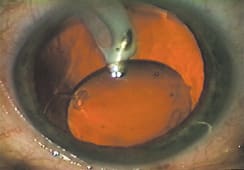
Healon5's high viscosity keeps the lens decentered long enough to let you place the I/A tip under the edge of the
IOL. |
|
Despite these advantages, one potential downside of Healon5 has caused even some seasoned surgeons to experience "high post-op IOP phobia:" the challenge of removing all of the Healon5 from behind the lens. Having been a clinical investigator in the Healon5 trials, and now having used it in more than 1,000 cases, I'd like to offer a few tips that should shorten the learning curve and help even the timid to go behind the lens and achieve total Healon5 removal.
Secrets to success
The two removal techniques recommended by Pharmacia during clinical trials were the "rock'n'roll" technique (Arshinoff) and the "two compartment technique" or "TCT" (Tetz). Because I had little experience going behind the IOL, the rock'n'roll was my technique of choice throughout the clinical trials.
This worked fine with the round-edged IOLs we used at that time. However, as I transitioned to truncated lenses to take advantage of lower posterior capsule opacification rates, I found that square-edged IOLs tended to act like "cookie cutters," trapping Healon5 between the posterior capsule and the IOL. This forced me to learn the two compartment technique.
|
|
|
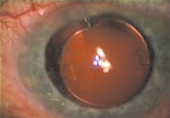
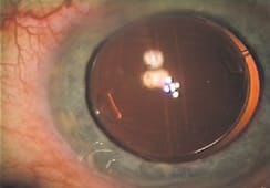
| Injecting BSS under the edge of the IOL "burps" out trapped Healon5. | The "Christie Crinkle" -- a slight vertical wrinkle in the capsule -- indicates that all the Healon5 has been evacuated. |
As I worked with this technique, I learned a number of key strategies:
- Don't center the IOL immediately after insertion. The high viscosity of Healon5 will allow the lens to remain decentered long enough to let you go under the edge of the IOL with the irrigation/aspiration (I/A) tip.
For years I went into the eye at this point in position 1 (for irrigation). However, the handpiece MUST be in position zero (no irrigation) in order to get under the IOL edge. - One "training wheels" tip when you're first attempting this: Inject a little more Healon5 under the edge of the IOL before going in with the I/A tip, particularly in small pupils or in cases where the lens seems uncomfortably close to the capsule. With vacuum set at 350 to 500 mm Hg and flow rates of at least 25 to 30 ml/min, the Healon5 will come right to the tip. Gently rotating the tip from right to left is the only movement you need to make.
- Occasionally, the IOL will slide under the I/A tip. When this happens, I resort to a rock'n'roll technique with a slight modification, which I call the "BSS burp." To execute the BSS burp I use a hydrodissection cannula to inject BSS under the edge of the
IOL. This "burps" the trapped Healon5 out from under the IOL and into the area anterior to the
IOL.
I often remove a surprising amount of Healon5 with the I/A tip after this maneuver -- certainly enough to cause an unwelcome IOP spike if it were left inside the eye. - When in doubt about trapped Healon5 I look for the "Christie Crinkle" -- a slight vertical wrinkle in the capsule. This wrinkle is a good indication that all the Healon5 has been evacuated.
Onward and upward
Healon5 has been my viscoelastic of choice for more than a year now, and I've learned to take advantage of the many attributes listed earlier. Meanwhile, meticulous removal using the two compartment technique has now become second nature to me and I've put "high post-op IOP phobia" behind me. Isn't that what ophthalmic surgery is all about?
Dr. Christie is founder of Scott & Christie and Associates, P.C., in Pittsburgh, and Good Looks Eyewear, an optical shop with two locations in the Pittsburgh area. He's been a principal investigator for several implant lens, viscoelastic and medication studies.
Making the Most of Ocucoat
By Paul Koch, M.D.
Ocucoat (2% methylcellulose) is a remarkably useful viscoelastic, for one simple reason: It changes its characteristics depending on how you apply it. Because of its unique physical properties it can behave like a semi-liquid gel, a dispersive viscoelastic or a cohesive viscoelastic. Injected one way it has a certain set of properties; injected another it behaves very differently.
Here are three ways you can use Ocucoat to make cataract surgery easier and safer.
Option one: a semi-liquid gel
If you place Ocucoat in an open space -- for example, on the surface of the cornea -- you'll see that it comes out as a semi-liquid, a soft substance that flows like fresh gelatin. It doesn't firm up and turn into a semi-solid; instead, it spreads out smoothly, guided by the pull of gravity.
In this form, Ocucoat is very useful for coating the cornea. As you know, the clarity of the view into the anterior chamber improves with surface moisture, making it easier to see the details and contours within the eye. Using BSS as the irrigant provides transient improvement, but using methylcellulose provides longer lasting clarity. A small amount of Ocucoat placed on the cornea spreads out, making a uniform, long-lasting coating over the corneal surface. Typically, one application will last an entire procedure, eliminating the need for periodically rinsing the cornea with BSS.
This improvement in clarity can also be used in another way. Usually a surgeon has no difficulty seeing the sideport incision, or at least remembering where it was made. Sometimes, however, the incision seals quickly and you may find that you poke at the cornea a few times without finding the opening. A small bit of Ocucoat placed on the periphery of the cornea improves your view of the stroma, letting you see the entire tract of the incision clearly, albeit for a brief moment. This will help you locate the incision quickly, safely, and without causing surface trauma.
Option two: a dispersive agent
When injecting a viscoelastic into the anterior chamber before capsulotomy, the idea is to use the viscoelastic as a dispersive agent and maintain the chamber. You generally want the viscoelastic to stay in one place as long as possible.
When Ocucoat is injected as a consistent, smooth single mass it behaves in exactly this manner; it maintains the space and provides a clear view of the ocular tissue. Yet it resists removal as a single substance; when you perform aspiration, it breaks up.
To get Ocucoat to behave this way, hold the cannula steady while you inject. Advance the cannula across the anterior chamber; then slowly withdraw while injecting. The Ocucoat is injected into itself, forming a smooth, uniform mass. In this form, it maintains the anterior chamber very well while providing excellent visibility. Leakage from the incision is usually modest and doesn't interfere with capsulotomy. (Naturally, as with any agent, if the eye softens, the capsulotomy can go off course. For that reason you should be liberal in using Ocucoat to maintain pressure in the anterior chamber.)
When Ocucoat is injected this way it lasts a long time. The vigorous irrigation and aspiration that take place during phacoemulsification don't easily remove the Ocucoat, which allows it to remain in protective contact with the endothelium. However, when injected this way for lens implantation, you'll find it difficult to remove it all at the end of the case because it will resist aspiration. A different technique is needed for this part of the procedure.
Option three: a cohesive agent
When performing capsulotomy you want to maintain the anterior chamber for as long as possible. In contrast, during implantation you only want to support the chamber for a few moments while the lens is injected. Once the lens is positioned you want the viscoelastic to come out of the eye as quickly as possible, and you want to leave as little in the eye as you can to avoid postoperative pressure problems. For this purpose you would typically use an agent that acts like a cohesive viscoelastic.
Ocucoat can also behave this way; all you have to do is change the way you inject it into the eye. To make it behave like a cohesive agent, place the cannula across the anterior chamber and slowly retract while injecting -- but this time wiggle the tip of the cannula back and forth. Try to generate a definite, visible squiggle of Ocucoat.
This thin ribbon of Ocucoat will have a very different clinical appearance than the uniform glob you created during capsulotomy. The method described earlier produces a smooth and uniform gel inside the eye; using this method you can see the outline of a continuous, narrow bead of viscoelastic.
In this state, Ocucoat is very cohesive. It has much better followability in the "squiggle" state than in the "glob" state. You can often irrigate it from the eye in a moment just by pressing against the incision with the I/A tip. Aspiration at the end of the case is typically rapid and efficient, leaving behind very little viscoelastic that might cause a postoperative pressure rise.
Making surgery easier
Using a viscoelastic with the ability to behave in three different ways can do a lot to simplify cataract surgery. And that's almost always a good thing, both for you and your patient.
Dr. Koch is chief medical editor for Ophthalmology Management and founder and director of Koch Eye Associates, with nine offices in Rhode Island and Connecticut. As a surgeon he's won numerous awards and helped to pioneer many advances in cataract surgery. Dr. Koch is a paid consultant to Bausch & Lomb.


