News
& Notes from the AAO Meeting
This month: glaucoma-related news. See the January issue for additional reports from the
meeting.
|
|
|
|
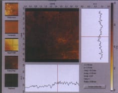
The HRT II Macular Edema Module detects clinically significant edema in a 0.5-mm radius circle around the fovea. |
|
HRT II now includes Macular Edema Module
Heidelberg Engineering added a Macular Edema Module to its Heidelberg Retina Tomograph (HRT) II. The new software package, available to current HRT II users for $7,000, uses topographic retinal thickness measurements and macular mapping techniques to objectively measure and allow clinicians to monitor diabetic and cystoid macular edema.
In "macula acquisition mode," the HRT II measures three-dimensional light reflectance from the retina and automatically scans up to 64 sequential images of the retina and aligns them to create a 3-D image in less than 5 seconds. The image measures 384 by 384 pixels and is composed of nearly 148,000 retinal thickness measurements. The examination doesn't require dilation.
Heidelberg says the new module makes the HRT II a valuable tool for retinal specialists as well as glaucoma specialists, and for general ophthalmologists, who can increase patient retention by avoiding unnecessary referrals.
GDx Access 3000 offers new capabilities and purchase options
Laser Diagnostic Technologies introduced the GDx Access 3000, which includes Custom Corneal Compensation capability. The Custom Corneal Compensator adjusts for each patient's corneal birefringence, making glaucoma exams more accurate, even for patients previously described as "corneal outliers." Also, an enhanced normative database allows comparison of the custom compensated images to normals.
The GDx Access 3000 now includes two features for image evaluation and patient monitoring that were previously available only in the original GDx Nerve Fiber Analyzer: a fundus image on the printout and the ability to view up to four exams on one printout.
In addition to the clinical improvements, LDT has made the GDx Access 3000 available on a new Ownership Plan and through its Freedom and Value monthly programs.
Zeiss Humphrey presents new fundus camera and new HFA test program
Zeiss Humphrey presented the new FF450plus telecentric fundus camera, which is designed especially for digital imaging. The FF450plus integrates with the VISUPAC Digital Image & Archive Management System.
A boost mode enhances detail in late-phase fluorescein angiography and ICG angiography when the FF450plus is used with the VISUPAC system. Ease of handling has been improved with the addition of automatic filter insertion via stepped motors and a smaller, more efficient control console.
Increased flash settings have been added, and a new, ergonomic table fosters better cable routing through access holes on either side of the camera. Also, an external cable arm can be used with VISUPAC-equipped systems. The camera adheres to the new ISO/FDIS 10940 standard, which stipulates criteria for patient exposure to light.
The company also introduced an exclusive kinetic test for the Humphrey Field Analyzer. The Enhanced Kinetic Program provides a full scope of Custom Kinetic testing, including kinetic tests in either step-by-step mode or an automatic mode; a selection of three pre-programmed tests with an option to use previously programmed meridians or to randomize the presentation of meridians; and the ability to add static points to a kinetic test, specify meridians by using a graphical interface, and print out results showing numeric values only.
Both the HFA II and the HFA II - i series instruments, models 750 and 750i, can run the Enhanced Kinetic Program.
|
|
|
|
The Selecta II is portable and can be attached to a slit lamp. |
Lumenis acquires HGM, upgrades Selecta
Lumenis Ltd. acquired HGM Medical Laser Systems Inc. of Salt Lake City, Utah. HGM has been marketing laser and delivery systems primarily to the ophthalmic market since the early 1980s.
Lumenis also introduced the Selecta II YAG laser for performing selective laser trabeculoplasty (SLT) to treat open-angle glaucoma. The Selecta II is a newly engineered system that uses advanced manufacturing technology and a flexible platform. It's portable and compact and can be attached to a slit lamp.
SLT lowers IOP by using short pulses of low-energy laser light to target specific melanin-containing cells in the trabecular meshwork, which stimulates an increase in fluid outflow. The selective technique is less traumatic to the eye than argon laser trabeculoplasty. Dr. Mark Latina reported that when SLT was used as primary therapy, it showed significant pressure reduction that was sustained over 36 months.
Update on blood flow and glaucoma
As part of the course "Frontiers in Ocular Blood Flow and Glaucoma: Principles and Practice," Dr. Ronald E. Frenkel addressed three key areas:
Evidence that increased IOP is not the sole cause of glaucoma. Current knowledge on this topic is reflected in the American Academy of Ophthalmology's new definition of glaucoma, which is no longer focused on IOP, and labels glaucoma a "a multifactorial optic neuropathy."
We now know that there is great individual variation in the susceptibility of the optic nerve to IOP-related damage, he said. For example:
- Of all patients who have increased IOP, only approximately 10% to 20% have glaucoma.
- Glaucoma progressed in 12% of the subjects in the Normal Tension Glaucoma Study whose IOP was controlled.
- Normal-tension glaucoma patients have microvascular, ischemic changes in the brain in and around the optic nerve.
- As Japanese patients age, their IOP decreases, but their incidence of glaucoma continues to increase with age. The prevalence of normal-tension glaucoma in Japan is three to four times that of primary open-angle glaucoma.
- An Australian study indicated that the strongest risk factor for glaucoma is a positive family history.
Evidence of vascular risk factors in glaucoma patients. The Normal Tension Glaucoma Study identified the two biggest risk factors for progression of visual field to be migraine headaches and disk hemorrhage. Also:
- Studies have shown that diastolic perfusion pressure below 55 mm Hg leads to increased risk of glaucoma damage. While this affects only 25% of cases, it raises the risk sixfold.
- Systemic hypertension is epidemiologically related to glaucoma, and vasospastic syndrome (generally in patients who have migraines or whose fingers and toes spasm after exposure to cold) is an established risk factor for glaucoma.
The interaction of IOP and blood flow. Dr. Frenkel explained that we know that elevated IOP increases resistance to blood flow into the eye, but we don't know whether glaucoma damage is caused directly, by the mechanical effects of high IOP, or whether increased IOP causes changes in hemodynamics. The result in either case, however, is apoptosis. Also:
- Both low blood pressure and high IOP reduce ocular perfusion pressure, and as perfusion pressure decreases, the glaucoma risk ratio increases.
- Patients with visual field progression also had dips in systolic, diastolic and mean arterial pressure.
- Visual field progression has also been associated with lower nighttime diastolic blood pressure. Therefore, nocturnal hypotension and a vulnerable optic nerve head may be a combination that causes glaucoma. However, patients who don't have glaucoma may also experience a blood pressure drop at night, but their bodies might autoregulate to compensate for that. This has led researchers to believe that most glaucoma patients could have normal perfusion but faulty autoregulation.
Dr. Frenkel said that at this point, it's almost proven that there is a vascular risk factor for glaucoma, and when that risk factor is in place, ischemia may play a central role in initiating apoptosis, which leads to retinal ganglion cell death.
He continued, "Our theories about the relationship between ocular blood flow and glaucoma have been around for a long time, but advances in technology are finally allowing us to test those theories.
"Our next step will be turning that knowledge into therapies. We've shown in joint studies with Dr. Latina that Trusopt increases blood flow, and other medications, such as Rescula and calcium channel blockers, are being studied. Now we need the necessary studies to prove that affecting blood flow will stop progressive glaucoma damage.
"Glaucoma has been so frustrating for us, as we've watched it progress in our patients whose pressures were controlled. But now that we're able to see beyond IOP, we may finally have something to offer those patients."
Unoprostone in patients on maximal medical therapy
Drs. Leslie S. Jones, L. Jay Katz and Undraa Altangerel conducted a compassionate protocol, retrospective, monocular study to determine the effectiveness of unoprostone 0.15% (Rescula) in lowering IOP in glaucoma patients who were receiving maximal medical therapy. "We were looking at how this medication performs in challenging cases," Dr. Jones said.
Maximal medical therapy was determined by Dr. Katz based on the number of medications patients could tolerate. Patients who had unoprostone added to both eyes and patients whose regimens underwent any other change at the time unoprostone was added were excluded from the study.
The 27 patients who were ultimately studied had advanced, progressing cases of glaucoma. Most were taking at least two medications; some were taking three or four. Seventy percent had undergone previous laser or incisional surgery; 67% of control eyes had a history of prior surgery.
The results showed:
- 63% of treated eyes experienced a pressure reduction
- 30% of treated eyes experienced a 20% reduction in IOP
- the average change in treated eyes was -2.4 mmHg (SD 4.9 mmHg)
- the average change in control eyes was -0.3 mmHg (SD 3.6 mmHg).
Thirty-seven percent of patients discontinued unoprostone due to insufficient IOP response or an adverse event; 15% underwent a filtering procedure; 48% remained on unoprostone.
"We were encouraged by the magnitude of IOP reduction in some of our difficult-to-treat patients," Dr. Jones said.
Brimonidine vs. latanoprost in progressive NTG
A group of researchers in Taiwan conducted a randomized, crossover study of changes in IOP, ocular perfusion pressure, blood pressure and heart rate in patients with progressive normal-tension glaucoma using either brimonidine (Alphagan) or latanoprost (Xalatan).
Twenty patients underwent 4 weeks each of washout I, medication I, washout II, medication II. The findings:
- IOP decreased by 3.0 mm Hg (21.3%) after latanoprost and by 1.4 mm Hg (10.1%) after brimonidine
- ocular perfusion pressure increased after latanoprost, not after brimonidine
- neither medication affected heart rate or blood pressure significantly.
Brimonidine tested in elderly patients
Until now, brimonidine (Alphagan) hadn't been evaluated as replacement therapy for topical beta blockers in a geriatric population.
As you know, geriatric glaucoma patients pose a challenge because they commonly suffer from co-morbid conditions, the symptoms of which often mirror the commonly reported systemic side effects of topical beta blocker therapy.
Dr. Robert J. Noecker et al. presented data from their prospective, multicenter, open-label crossover evaluation of 89 patients who were at least 65 years old and using topical beta blocker therapy for glaucoma for at least 6 months.
Patients who had used brimonidine before, had intraocular or laser surgery within the past 3 months, were using systemic beta blockers, or had inflammatory glaucoma, a history of steroid glaucoma or a contraindication to brimonidine were excluded.
The patients were washed out from their topical beta blockers for 1 month, and then instilled brimonidine b.i.d. for 1 month. After that month:
- the mean reduction in IOP at peak drug effect was 1.4 ± 0.4 mm Hg (8.2%)
- there was a mean improvement in forced expiratory volume with brimonidine, compared with topical beta blocker therapy, of 26.2 liters/minute
- a mean increase in cardiac function of 2.3 beats per minute was noted.

Medications in black and nonblack patients
Dr. Peter A. Netland presented data from a 12-month, planned, prospective study comparing the IOP-lowering effect of travoprost, latanoprost and timolol in black (N = 177) and nonblack (N = 610) patients.
Key findings included that the IOP lowering effect of travoprost was equal to or superior to latanoprost, and superior to that of timolol 0.5%. Also, travoprost was significantly more effective than either latanoprost or timolol in reducing IOP in black patients. Specifically, in black patients, travoprost lowered mean IOP more than timolol or latanoprost by up to 4.6 mm Hg and 2.4 mm Hg respectively.
Dr. Netland also covered medication responder rates. (Responders were defined as patients on treatment with an IOP drop of 30% or greater, or a final intraocular pressure of 17 mm Hg or less.) Travoprost had the highest responder rate at 54.7%, followed by latanoprost (49.6%) and timolol (39.0%).
Dr Netland noted that travoprost was found to be safe and well tolerated, and represents an excellent addition to therapeutic glaucoma management options.
A new medication in practice
Dr. Thomas Walters, who helped develop brimonidine 0.15% (Alphagan P), the new formulation of brimonidine 0.2% (Alphagan), talked with Ophthalmology Management about his experience with the new medication. "The study findings have borne out in my practice," he said.
He explained that Alphagan P was an attempt to improve on the original formulation in regard to ocular allergy rate and the less significant issues of patient dry mouth and drowsiness. The result was a medication that demonstrated comparable efficacy to Alphagan, with the lowest effective dose of active drug, but with 41% less incidence of ocular allergy. Alphagan P is preserved with Purite, the same preservative used in Refresh Tears lubricant eye drops.
"This was really a case of taking a good product and making it better," Dr. Walters said.
Trials of new glaucoma medication advance
Santen has begun the U.S. Phase II clinical trial of DE-085 (AFP-168), a new prostaglandin-based therapeutic agent for glaucoma. The trial will verify safe, effective dosage and administration in a small number of patients. DE-085 was developed in cooperation with Asahi Glass Co., Ltd.
Fixed combination vs. concomitant therapy
Dr. Michael Ehrenhaus et al. conducted a retrospective review of 20 patient charts to compare the IOP-lowering effect of fixed-combination dorzolamide 2% and timolol 0.5% (Cosopt) to concomitant treatment with the medications. Baseline IOPs at two visits before switching medications were 18 mm Hg and 18.05 mm Hg respectively.
Based on the data after the switch to the fixed combination, which included an additional 1.25 mm Hg IOP decrease (to a mean of 16.75 mmHg), the doctors concluded that switching to a fixed combination of dorzolamide and timolol from concomitant therapy produces a clinically significant reduction in IOP.
3-month comparison of bimatoprost and timolol/dorzolamide
In a 3-month study, Dr. Anne L. Coleman et al. compared the IOP-lowering efficacy and safety of bimatoprost (Lumigan) q.d. compared with timolol/dorzolamide (Cosopt) b.i.d.
The study, involving 177 ocular hypertension/chronic glaucoma patients, was prospective, multicenter, randomized and investigator-masked. The inclusion criteria were:
- at least 21 years of age
- ocular hypertension or glaucoma requiring bilateral treatment
- BCVA of 20/100 or better in each eye
- IOP between 22 mm Hg and 34 mm Hg in at least one eye at baseline at 8 a.m. after at least 2 weeks of beta blocker monotherapy.
Six visits were scheduled: prestudy, baseline, week 1, and months 1, 2, and 3. IOP was measured at each visit at 8 a.m. and 10 a.m. It was also measured at the baseline and 3-month visits at 2 p.m., 4 p.m. and 8 p.m.
Efficacy was analyzed using the eye with the highest IOP determined at baseline at 8 a.m. Based on their data, the researchers reported:
- bimatoprost provided greater IOP lowering (0.5 mm Hg to 3.2 mm Hg) than timolol/dorzolamide at all follow-up visits at all time points
- at month 3 at 8 a.m., a significantly greater percentage of patients reached lower target IOPs of less than or equal to 13, 14, 15 and 16 mm Hg with bimatoprost than with timolol/dorzolamide (Almost one-third of the bimatoprost patients achieved IOP less than or equal to 16 mm Hg.)
- conjunctival hyperemia was more common with bimatoprost; taste perversion and ocular burning and stinging were more common with timolol/dorzolamide. (Only 3% of patients in each treatment group discontinued because of adverse events.)
The study indicates that it's possible to further lower IOP with monotherapy, without the combination-therapy risks of more side effects, including potentially serious cardiovascular ones.


