Case One
Pigment dispersion syndrome and refractive patients
Recently, I've seen several patients with significant glaucoma following refractive procedures. In nearly all of these patients, signs suggested preoperatively that trouble could be coming.
Patient history/presentation: A 46-year-old white male was referred for glaucoma after having undergone LASIK 1 year prior. The glaucoma developed after the LASIK had been performed. The pressure rise occurred during the postoperative period when the patient was placed on steroids. Even after the cessation of steroids, the IOP continued to be too high. There were multiple attempts to control the intraocular pressure with medications.
Examination/findings: Vision was 20/20 OU. Goldmann tonometry readings were 25 mm Hg and 17 mm Hg. The external exam was within normal limits. There was early afferent pupillary defect OD. Pachymetry showed corneal thicknesses of 512 microns and 507 microns. The slit-lamp exam revealed quiet conjunctiva; a faint LASIK scar was present; the iris was flat without transillumination defects; and the lens was clear.
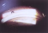
Gonioscopy revealed a deep angle with heavy pigmentation of the trabecular meshwork region. After dilation, the lens revealed a posterior pigment line and an asymmetric cup/disc ratio. The right eye had a 0.8 C/D, and the left eye had a 0.4 C/D. Visual fields revealed an arcuate defect in the right eye and a normal left eye. The findings are consistent with pigment dispersion syndrome (PDS).
Treatment: Multiple medication changes and ALT failed to lower the IOP. A trabeculectomy was required for IOP control. Severe visual fluctuation in the vision has occurred postoperatively with substantial hyperopia being present most of the time. Multiple attempts to balance the vision have resulted in multiple pairs of spectacles used during the day to allow for acceptable vision.
Discussion: Pigment dispersion syndrome is most common in young myopic males. These individuals are also those who often want LASIK or other refractive procedures. The classic findings with pigment dispersion syndrome are a Krukenberg's spindle on the corneal endothelium, iris transillumination defects, trabecular meshwork pigmentation, and pigment on the posterior lens surface (Vossius ring).
Unfortunately, not all the findings are consistently present for patients with pigment dispersion. For patients with early trabecular obstruction, there can be a tendency toward significant IOP swings or IOP asymmetry. It's not commonplace, however, to check diurnal curves on refractive surgery candidates. So how do we try to identify patients who may have undiagnosed dispersion and may get into trouble after refractive surgery?
To find the dispersion patient who could spell trouble:
- Review the records from prior treating doctors, looking for occasional higher IOP, especially in one eye.
- Perform gonioscopy on your patients and look for trabecular meshwork pigment that is heavier in one eye than in the other.
- Check for classic Krukenberg's spindles or transillumination defects in the iris.
- Look for slightly asymmetric IOP or cups.
- Look for a pigment line on the posterior surface of the lens.
- Ask whether the patient has noted blurred vision in one or both eye(s) after activities such as aerobic classes or skiing.
Even the nondispersion patient can be a steroid responder; therefore, as a rule, I tell patients placed on steroid eye drops that they could develop an IOP rise and they need to specifically have their IOP checked. This is especially the case in those patients you co-manage and when there may be a tendency for the doctor seeing the patient to not want to check the IOP because the patient just had LASIK.
To manage refractive candidates who do have PDS:
- Consider a diurnal curve.
- Consider a provocative test with steroids. It seems that the steroid response is a significant factor contributing to a rise in the IOP in most of the patients after LASIK. However, most of them had something else (such as PDS) that also contributed to the increased IOP.
- Discuss the PDS with the patient and the possibility of glaucoma developing in the future.
- Try to select the procedure with the least likelihood of requiring prolonged steroids.
Should refractive procedures be done on glaucoma suspects or patients with glaucoma? This has been a big debate. Unfortunately, the human body responds differently than we would like it to at times. When you speak with your patients preoperatively, and there is a possibility of post-op glaucoma, don't minimize this as an issue. Explain the risk thoroughly. Otherwise, if they do develop glaucoma, the feel as if they've been lied to.
Case One was submitted by E. Randy Craven, M.D., from Glaucoma Consultants of Colorado in Littleton, Colo. If you'd like to comment on this case, e-mail Ophthalmology Management at ifftda@boucher1.com.
|
|
|
|
Gonioscopy showed increased
trabecular
pigmentation in both eyes. The right eye
is pictured. |
|
Case Two
"Burnt-out" pigmentary glaucoma
Patient history/presentation: A 50-year-old white male presented for a second opinion. He had been diagnosed with normal-tension glaucoma 2 years previously during a routine eye exam. His IOP was in the low teens in both eyes, but he had a glaucomatous visual field defect and glaucomatous optic nerve damage in his right eye. He had been placed on Xalatan once daily in each eye, but discontinued it on his own shortly after that examination. He had worn glasses for myopia for many years.
|
|
|
|
A Humphrey 24-2 threshold visual field test showed a defect
OD. |
Examination/findings: On examination, visual acuity was 20/20 in each eye with correction. The applanation tension was 14 mm Hg OD and 12 mm Hg OS, on no glaucoma medication. Slit-lamp examination demonstrated mild corneal endothelial pigment dusting but no Krukenberg's spindle. There were a few peripheral radial iris transillumination defects inferiorly in both eyes.
A Humphrey 24-2 threshold visual field test was normal in the left eye, and showed a superior arcuate defect in the right eye, unchanged from 2 years previously. Gonioscopy showed increased trabecular pigmentation in both eyes. Dilated fundus examination was normal in both eyes except for glaucomatous optic nerve damage in the right eye. There were no disc hemorrhages in either eye.
Diagnosis: Nonprogressive normal-tension glaucoma because of former pigmentary glaucoma.
Treatment: It was decided to not treat this patient. He is being examined annually to be certain of a lack of progression or future increase in IOP.
|
|
|
|
Glaucomatous optic nerve damage was detected in the right eye. |
|
Discussion: Pigmentary glaucoma is most commonly seen in young myopic males and less commonly in females. Pigment dispersion is greatest from about age 20 to 45 years. It is believed to be caused by a rubbing off of posterior iris pigment by zonules of myopes with a posterior iris convexity. As with all glaucomas, the visual loss may be asymptomatic and not be discovered until later in life when a visual field defect is found or optic disc damage is seen by the ophthalmologist.
For these patients, if IOP is normal, usually no treatment is necessary. Stereo disc photography can help conclusively demonstrate a lack of progression of "burnt-out" pigmentary glaucoma.
Case Two was submitted by Edward J. Rockwood, M.D., from the Cole Eye Institute at the Cleveland Clinic. If you'd like to comment on this case, e-mail Ophthalmology Management at ifftda@boucher1.com.


