Wavefront Deluxe
Combining wavefront technology, topography and autorefractometry in a powerful new diagnostic tool.
BY CHRISTOPHER KENT,
SENIOR ASSOCIATE EDITOR
Now that wavefront technology is becoming a reality in the United States, ophthalmologists are beginning to look more closely at the issues raised by this new type of diagnostic tool. Which version of the technology works best? How can we maximize its advantages? How can we integrate it most effectively with other diagnostic instruments?
The OPD Scan from Nidek, which received FDA 510(k) clearance in April, offers some potential answers. The OPD Scan combines three technologies in a single instrument: dynamic skiascopy (wavefront) technology, placido disk corneal topography and autorefractometry -- and it measures your patient's eye with all three simultaneously. It then creates a series of refractive power maps that show the total optical system aberrations in the eye. (OPD, incidentally, stands for optical path difference.)
Riding the wave
As you know, wavefront technology uses reflected light to measure refractive power at hundreds of points across the eye. Unlike Hartmann-Shack wavefront technology, which uses a lenslet array to measure how far the reflected light has been displaced, dynamic skiascopy measures the timing of the reflected light.
|
|
|
|
Nidek's OPD Scan avoids alignment problems by taking three measurements simultaneously. |
|
According to Nidek, this eliminates a potential problem: Unusual corneal shapes can make it difficult for Hart-mann-Shack instruments to obtain measurements. (In an informal, comparative test conducted at ASCRS in which a patient with keratoconus was scanned, a machine using the Hartmann-Shack method and another using Tscherning-type aberrometry had problems. The OPD Scan did not.)
The OPD Scan's projecting and receiving systems rotate around the optical axis, measuring refractive power at every 1° meridian. At the same time, the OPD Scan measures corneal topography and performs standard autorefraction.
Data presentation
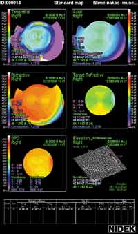
Once the data is collected, it can be combined and displayed in any of 12 different diagnostic map formats. (Six of these maps can be shown onscreen at the same time.)
Map formats available include:
- axial map
- elevation map
- instantaneous or tangential map
- refractive power map, showing the degree of spherical aberration (calculated according to Snell's Law)
- the "OPD map," which displays the refractive error of the visual system within a 6-mm zone centered around the visual axis. It also indicates the average refractive power at 1.5 and 2.5-mm radii from the visual axis, to give you a sense of the shape of the cornea and the impact of aberrations as you move away from the center.
- wavefront higher-order aberration map, showing only third-order or higher aberrations
- wavefront total aberration map
- refractive power map, integrating topography and wavefront analysis data, presented in the form of a traditional topographic map
- target refractive map, showing the estimated corneal refractive power to achieve emmetropia postoperatively
- Zernike graph, showing the contribution of each type of aberration
- photokeratoscope
- difference map.
You can move the cursor across any of these maps and see what the refractive power is at any given point. You can also print them out using an optional color printer. (A built-in thermal printer is provided for printing the autorefraction and autokeratometry data.)
Features and benefits
Practical advantages of the OPD Scan include:
|
|
|
|
The OPD Scan allows you to display six different diagnostic maps onscreen simultaneously. |
- It completes its measurements in less than half a second.
- The data is high resolution. The OPD scans 1,440 measurement points for aberration and autorefractor data, and 6,480 points for corneal topography data.
- The OPD Scan can measure very large refractive ranges -- from -20 to +22 diopters of spherical error, and up to 12 diopters of cylinder. (Other wavefront technologies are more limited.)
- Because all measurements are taken at once using a single instrument, alignment is automatic and registration of the data is more accurate.
- Dry eye doesn't affect the measurement significantly because the OPD Scan uses infrared light.
- Because data is gathered and combined internally, there's no need to track different pieces of paper and information, which can be time-consuming and increase the risks of human error.
Using the OPD Scan in practice
Howard Gimbel, M.D., of Calgary, Alberta, Canada, has worked with the OPD Scan for nearly a year and a half. "Wavefront technology is essential in today's refractive surgery practice," he observes. "It gives you a lot more information about the eye than previous technologies. And because the OPD combines this with placido disk mapping and standard autorefraction, all with the same registration, we don't have to worry about head and eye position or fixation. It's an ideal situation.
"Also, it's easy to use -- much like an autorefractor or topographer. I think the OPD Scan is a good investment."
Arturo Chayet, M.D., who practices in Tijuana, Mexico, uses the OPD Scan for both surgery and research. "The OPD Scan is very compact, ergonomic and easy to use. I particularly like that it provides a map giving you the power of the visual system at each point, and the convenience of a touchscreen for data entry. Also, the topography system is very good, even used by itself. It's definitely worth the cost if you're looking for wavefront analysis."
Custom ablation using "Final Fit"
Nidek is developing a compatible software system that will interface the OPD Scan with Nidek's EC-5000 Excimer Laser System to produce custom ablations.
Dr. Gimbel has been using a prototype of the Final Fit system for more than a year. "The first patient we treated using the Final Fit system had a decentered ablation from a previous procedure. Although his refraction was 20/20, his quality of vision wasn't satisfactory. He complained of ghosting, and his eyes had difficulty working together.
"We were conservative in our use of the OPD Scan and laser because these were prototypes. Nevertheless, the patient experienced considerable subjective improvement in his vision. Since then, we've offered to do more refinement, but he's content with that first result."
Bringing it all together
With three state-of-the-art technologies integrated into a single device, the OPD Scan clearly has a lot to offer. For more information, call Nidek at (800) 223-9044 or visit their Web site at www.nidek.com.
Are you aware of new products or technology that have made (or are likely to make) a significant difference in practice? Contact Christopher Kent at kentcx@boucher1.com to find out about possible coverage in a future issue.


